

Attenuation of myocardial injury in mice with functional deletion of the circadian rhythm gene mPer2
- PMID: 20061537
- PMCID: PMC2838551
- DOI: 10.1152/ajpheart.01280.2008
Attenuation of myocardial injury in mice with functional deletion of the circadian rhythm gene mPer2
Abstract
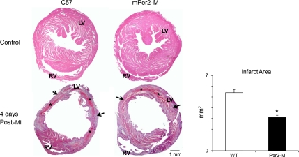
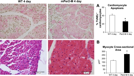
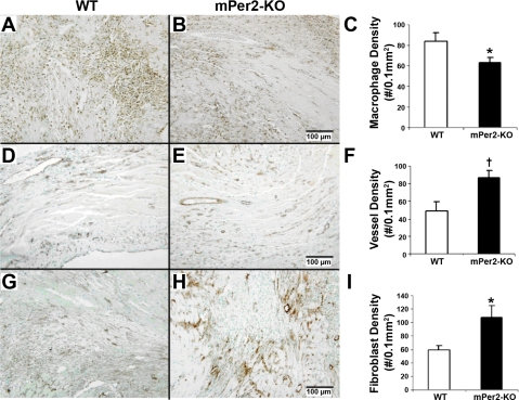
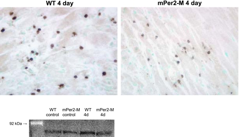
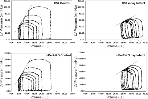
Variations in circadian rhythms are evident in the incidence of cardiovascular disease, and the risk of cardiovascular events increases when rhythms are disrupted. The suprachiasmatic nucleus is the central circadian pacemaker that regulates the daily rhythm of peripheral organs. Diurnal rhythms have more recently been shown to exist in myocardial tissue and are involved in metabolism and contractile function. Thus we sought to determine whether the functional deletion of the circadian rhythm mouse periodic gene 2 (mPer2) would protect the heart against ischemic injury. Nonreperfused myocardial infarction was induced in anesthetized, ventilated C57 (n = 17) and mPer2 mutant (mPer2-M; n = 15) mice via permanent ligation of the left anterior descending coronary artery. At 4 days post-myocardial infarction, we observed a 43% reduction of infarct area in mPer2-M mice compared with wild-type mice. This is coincident with 25% less macrophage infiltration, 43% higher capillary density, 17% increase in hypertrophy, and 15% less cardiomyocyte apoptosis in the infarct zone. Also, matrix metalloproteinase-9 was expressed in inflammatory cells in both groups, but total protein was 40% higher in wild-type mice, whereas it was not elevated in mPer2-M mice in response to injury. The functional deletion of the mPer2 gene reduces the severity of myocardial infarct injury by limiting the inflammatory response, reducing apoptosis, and inducing cardiomyocyte hypertrophy, thus preserving cardiac function. These findings collectively imply that the disruption of the circadian clock gene mPer2 is protective. Understanding the interactions between circadian rhythm genes and cardiovascular disease may provide insights into potential preventative and therapeutic strategies for susceptible populations.
Figures
Similar articles
-
Cardioprotection via preserved mitochondrial structure and function in the mPer2-mutant mouse myocardium.Am J Physiol Heart Circ Physiol. 2013 Aug 15;305(4):H477-83. doi: 10.1152/ajpheart.00914.2012. Epub 2013 Jun 14. Am J Physiol Heart Circ Physiol. 2013. PMID: 23771689 Free PMC article.
-
A Novel Diabetic Mouse Model for Real-Time Monitoring of Clock Gene Oscillation and Blood Pressure Circadian Rhythm.J Biol Rhythms. 2019 Feb;34(1):51-68. doi: 10.1177/0748730418803719. Epub 2018 Oct 2. J Biol Rhythms. 2019. PMID: 30278816 Free PMC article.
-
Short-term disruption of diurnal rhythms after murine myocardial infarction adversely affects long-term myocardial structure and function.Circ Res. 2014 May 23;114(11):1713-22. doi: 10.1161/CIRCRESAHA.114.302995. Epub 2014 Mar 31. Circ Res. 2014. PMID: 24687134
-
Cardiac period 2 in myocardial ischemia: clinical implications of a light dependent protein.Int J Biochem Cell Biol. 2013 Mar;45(3):667-71. doi: 10.1016/j.biocel.2012.12.022. Epub 2013 Jan 3. Int J Biochem Cell Biol. 2013. PMID: 23291353 Free PMC article. Review.
-
Effect of KLF15-Mediated Circadian Rhythm on Myocardial Infarction: A Narrative Review.Int J Mol Sci. 2025 May 18;26(10):4831. doi: 10.3390/ijms26104831. Int J Mol Sci. 2025. PMID: 40429972 Free PMC article. Review.
Cited by
-
The Impact of the Circadian Genes CLOCK and ARNTL on Myocardial Infarction.J Clin Med. 2020 Feb 10;9(2):484. doi: 10.3390/jcm9020484. J Clin Med. 2020. PMID: 32050674 Free PMC article.
-
Coordination of cardiac rhythmic output and circadian metabolic regulation in the heart.Cell Mol Life Sci. 2018 Feb;75(3):403-416. doi: 10.1007/s00018-017-2606-x. Epub 2017 Aug 21. Cell Mol Life Sci. 2018. PMID: 28825119 Free PMC article. Review.
-
The role of sulfur dioxide in the regulation of mitochondrion-related cardiomyocyte apoptosis in rats with isopropylarterenol-induced myocardial injury.Int J Mol Sci. 2013 May 21;14(5):10465-82. doi: 10.3390/ijms140510465. Int J Mol Sci. 2013. PMID: 23698774 Free PMC article.
-
The Cardiac Circadian Clock: Implications for Cardiovascular Disease and its Treatment.JACC Basic Transl Sci. 2023 Jun 21;8(12):1613-1628. doi: 10.1016/j.jacbts.2023.03.024. eCollection 2023 Dec. JACC Basic Transl Sci. 2023. PMID: 38205356 Free PMC article. Review.
-
Coronary artery ligation and intramyocardial injection in a murine model of infarction.J Vis Exp. 2011 Jun 7;(52):2581. doi: 10.3791/2581. J Vis Exp. 2011. PMID: 21673649 Free PMC article.
References
-
- Arjona A, Sarkar DK. The circadian gene mPer2 regulates the daily rhythm of IFN-gamma. J Interferon Cytokine Res 26: 645–649, 2006 - PubMed
-
- Bourin P, Ledain AF, Beau J, Mille D, Levi F. In-vitro circadian rhythm of murine bone marrow progenitor production. Chronobiol Int 19: 57–67, 2002 - PubMed
-
- Bray MS, Shaw CA, Moore MW, Garcia RA, Zanquetta MM, Durgan DJ, Jeong WJ, Tsai JY, Bugger H, Zhang D, Rohrwasser A, Rennison JH, Dyck JR, Litwin SE, Hardin PE, Chow CW, Chandler MP, Abel ED, Young ME. Disruption of the circadian clock within the cardiomyocyte influences myocardial contractile function, metabolism, and gene expression. Am J Physiol Heart Circ Physiol 294: H1036–H1047, 2008 - PubMed
-
- Corwin JV, Vargo JM. Light deprivation produces accelerated behavioral recovery of function from neglect produced by unilateral medial agranular prefrontal cortex lesions in rats. Behav Brain Res 56: 187–196, 1993 - PubMed
-
- Dardente H, Cermakian N. How many pieces to build a circadian clock? [In French.] Med Sci (Paris) 21: 66–72, 2005 - PubMed
Publication types
MeSH terms
Substances
Grants and funding
LinkOut - more resources
Full Text Sources
Other Literature Sources
Medical
Molecular Biology Databases