. 2010 Jan;22(1):108-23.
doi: 10.1105/tpc.109.065490. Epub 2010 Jan 8.

Arabidopsis CULLIN4-damaged DNA binding protein 1 interacts with CONSTITUTIVELY PHOTOMORPHOGENIC1-SUPPRESSOR OF PHYA complexes to regulate photomorphogenesis and flowering time

Affiliations

Arabidopsis CULLIN4-damaged DNA binding protein 1 interacts with CONSTITUTIVELY PHOTOMORPHOGENIC1-SUPPRESSOR OF PHYA complexes to regulate photomorphogenesis and flowering time

Haodong Chen et al. Plant Cell. 2010 Jan.

Abstract

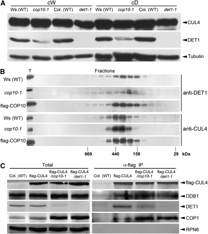
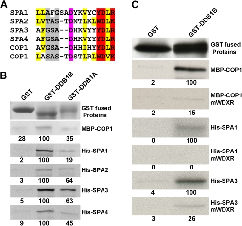
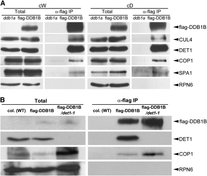
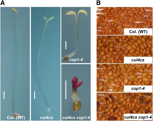
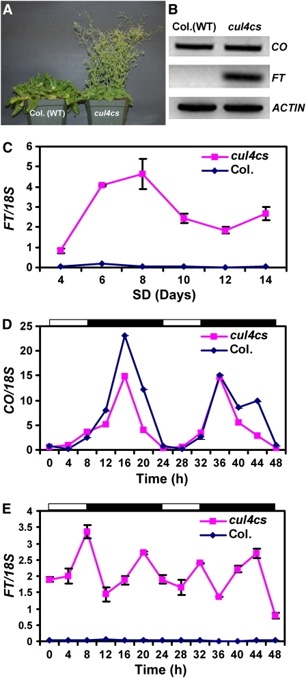
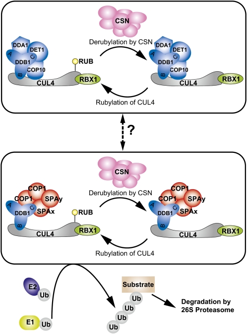
CONSTITUTIVELY PHOTOMORPHOGENIC1 (COP1) possesses E3 ligase activity and promotes degradation of key factors involved in the light regulation of plant development. The finding that CULLIN4 (CUL4)-Damaged DNA Binding Protein1 (DDB1) interacts with DDB1 binding WD40 (DWD) proteins to act as E3 ligases implied that CUL4-DDB1 may associate with COP1-SUPPRESSOR OF PHYA (SPA) protein complexes, since COP1 and SPAs are DWD proteins. Here, we demonstrate that CUL4-DDB1 physically associates with COP1-SPA complexes in vitro and in vivo, likely via direct interaction of DDB1 with COP1 and SPAs. The interactions between DDB1 and COP1, SPA1, and SPA3 were disrupted by mutations in the WDXR motifs of MBP-COP1, His-SPA1, and His-SPA3. CUL4 cosuppression mutants enhanced weak cop1 photomorphogenesis and flowered early under short days. Early flowering of short day-grown cul4 mutants correlated with increased FLOWERING LOCUS T transcript levels, whereas CONSTANS transcript levels were not altered. De-etiolated1 and COP1 can bind DDB1 and may work with CUL4-DDB1 in distinct complexes, but they mediate photomorphogenesis in concert. Thus, a series of CUL4-DDB1-COP1-SPA E3 ligase complexes may mediate the repression of photomorphogenesis and, possibly, of flowering time.


Figures

Figure 1.
Figure 1.
CUL4 and COP1 Interact with Each Other in the Absence of the CDD Complex. (A) The DET1 protein level is dramatically reduced in cop10-1 mutants. Total protein extracts from 6-d-old continuous white light–grown (cW) and dark-grown (cD) seedlings were examined by protein gel blot analysis using anti-CUL4, anti-DET1, and anti-Tubulin. Tubulin protein level was used as a sample loading control. (B) Protein gel blot analyses showing the gel filtration patterns of DET1 and CUL4 proteins in Ws (wild type), cop10-1, and flag-COP10 (in cop10-1 background), respectively. Molecular masses are indicated at bottom. T, total unfractionated extracts. (C) Flag-CUL4 associates with DDB1 and COP1 in vivo in wild-type, cop10-1, and det1-1 backgrounds. Total protein extracts prepared from 6-d-old dark-grown wild-type, 35S:flag-CUL4, 35S:flag-CUL4/cop10-1, and 35S:flag-CUL4/det1-1 transgenic Arabidopsis seedlings were incubated with anti-flag antibody-conjugated agarose ( α -flag). The precipitates and total extracts were subjected to immunoblot analysis with antibodies against flag, DDB1, DET1, COP1, and RPN6.
Figure 2.
Figure 2.
DDB1A and DDB1B Interact with COP1 and SPA Proteins in Vitro. (A) Alignment of the WDXR regions of COP1, SPA1, SPA2, SPA3, and SPA4. The WDXR motif is highlighted in red. Pink, conserved D or E; yellow, hydrophobic amino acids; gray, small amino acids. (B) In vitro coimmunoprecipitation of COP1 and SPA proteins by recombinant DDB1A and DDB1B. Recombinant MBP-COP1 purified from Escherichia coli and 35S-labeled His-SPA1, His-SPA2, His-SPA3, and His-SPA4 generated by the TNT system were used as prey molecules and incubated with unlabeled GST, GST-DDB1A, and GST-DDB1B, respectively. Anti-GST antibody fused to agarose was used for immunoprecipitation. Pellet fractions were resolved by SDS-PAGE and visualized by Coomassie blue staining (for GST fused proteins), anti-COP1 immunoblot (for MBP-COP1), and autoradiography using a phosphor imager (for SPA proteins). Quantification was based on normalization against the value of the background region. Band intensities of MBP-COP1, His-SPA1, His-SPA2, His-SPA3, and His-SPA4 pulled down by GST-DDB1B were each set to 100. Relative band intensities were then calculated and are indicated by numbers below blots. (C) Point mutations within the WDXR motifs of COP1 and SPA proteins disrupt their interactions with DDB1. MBP-COP1mWDXR contains the following mutations in the WDXR motifs: D534A, R536A, D576A, and K578A; His-SPA1mWDXR contains mutations D879A and R881A; His-SPA3mWDXR contains mutations D689A and R691A. Wild-type and mutated recombinant MBP-COP1 purified from E. coli, and wild-type and mutated 35S-labeled His-SPA1, His-SPA3 generated by the TNT system were used as prey molecules and incubated with GST and GST-DDB1B, respectively. Anti-GST antibody fused to agarose was used for immunoprecipitation. Pellet fractions were resolved by SDS-PAGE and visualized by anti-GST immunoblot (for GST fused proteins), anti-MBP immunoblot (for wild-type and mutated MBP-COP1), and autoradiography using x-ray films (for wild-type and mutated SPA proteins). Quantification was performed for each pair of wild-type and mutated prey proteins. Band intensities of MBP-COP1, His-SPA1, and His-SPA3 pulled down by GST-DDB1B were set to 100. Relative band intensities were then calculated and are indicated by numbers below blots.
Figure 3.
Figure 3.
CUL4-DDB1 Interacts with the COP1-SPA Complexes in Vivo. (A) Flag-DDB1B associates with CUL4, DET1, COP1, and SPA1 in vivo. Total protein extracts prepared from 6-d-old continuous white light–grown (cW) and dark-grown (cD) wild-type and 35S:flag-DDB1B/ddb1a (abbreviated as flag-DDB1B) transgenic Arabidopsis seedlings were incubated with anti-flag antibody-conjugated agarose ( α -flag). The precipitates and total extracts were subjected to immunoblot analysis with antibodies against flag, CUL4, DET1, COP1, SPA1, and RPN6. RPN6 was used as a control. (B) Flag-DDB1B associates with COP1 in wild-type and det1-1 backgrounds. Total protein extracts prepared from 6-d-old dark-grown wild-type, 35S:flag-DDB1B/ddb1a (abbreviated to be flag-DDB1B), and 35S:flag-DDB1B/det1-1 transgenic Arabidopsis seedlings were incubated with anti-flag antibody-conjugated agarose ( α -flag). The precipitates and total extracts were subjected to immunoblot analysis with antibodies against flag, DET1, COP1, and RPN6. RPN6 was used as a control.
Figure 4.
Figure 4.
CUL4 and COP1 Function Together to Regulate Photomorphogenesis. (A) Repression of CUL4 enhances the seedling phenotype of the weak cop1-4 allele in the dark. Different Arabidopsis lines (labeled at bottom) were grown in complete darkness for 6 d. Bars = 1 mm. (B) Repression of CUL4 increases anthocyanin accumulation in cop1-4 seeds. The cul4cs and cop1-4 seeds are homozygous, while the cul4cs cop1-4 are F2 seeds from hybrid F1 plants.
Figure 5.
Figure 5.
CUL4 Is Involved in the Regulation of Flowering Time under SDs. (A) cul4cs flowers early in SDs. cul4cs and its wild-type counterpart Col were grown for 90 d under SDs (8 h light/16 h dark). (B) Levels of CO and FT transcripts in cul4cs and wild-type Col. Total RNA samples were collected from 20-d-old Col and cul4cs plants grown under SDs (8 h light/16 h dark) and harvested at ZT 16. mRNA levels of CO, FT, and ACTIN2 were tested by RT-PCR with three repeats. ACTIN2 transcripts were used as control. (C) FT transcription starts very early in SD-grown cul4cs. Total RNA samples were collected from 4-, 6-, 8-, 10-, 12-, and 14-d-old Col and cul4cs plants grown under SDs (8 h light/16 h dark) and harvested at ZT 16. Levels of FT mRNA and 18S rRNA were tested by real-time PCR. Relative amounts of FT transcripts were normalized to the levels of 18S rRNA in the same sample. (D) and (E) Circadian patterns of CO (D) and FT (E) mRNA levels in cul4cs and wild-type Col. The plants were grown under SDs (8 h light [white bar]/16 h dark [black bar]) for 18 d and then RNA samples were collected every 4 h, at the times shown, after dawn. Levels of CO and FT mRNA and 18S rRNA were analyzed by real-time PCR. Relative amounts of CO and FT transcripts were normalized to the levels of 18S rRNA. For (C) to (E), mean and sd values of four replicates are shown. Some error bars are too small to be shown in the figure.
Figure 6.
Figure 6.
Effects of Point Mutations on the Surface of the DDB1 Protein. (A) Structural diagram depicting the locations of the three groups of mutations on the surface of DDB1. Mutated residues are indicated. (B) Effects of DDB1 mutations on binding to CUL4 and COP1. Wild-type or point-mutated DDB1B coding sequences fused to the flag peptide and driven by the 35S promoter were transformed into ddb1a mutants, and the transgenic plants were used for coimmunoprecipitation. DDB1B, mBPA, and mBPB indicate wild-type flag-DDB1B, flag-DDB1B with point mutations within the BPA domain, and flag-DDB1B with point mutations within the BPB domain, respectively. Total protein extracts from 6-d-old light-grown Arabidopsis seedlings were incubated with anti-flag antibody-conjugated agarose ( α -flag). The precipitates and total extracts were subjected to immunoblot analysis with antibodies against flag, CUL4, and COP1. (C) Phenotypes of 38-d-old ddb1a and mBPC plants under LDs (16 h light/8 h dark). The ddb1a mutant is in the left pot, and the mBPC mutant is in the right pot. mBPC contains flag-DDB1B with point mutations in the BPC domain transformed into the ddb1a mutant background. (D) DDB1 transcript levels in ddb1a and mBPC. Total RNA samples were collected from 4-week-old ddb1a and mBPC plants, and DDB1B expression levels were quantified by RT-PCR with three repeats. ACTIN2 transcripts were used as control. (E) DDB1 protein levels in ddb1a and mBPC. Total proteins were extracted from 15-d-old ddb1a and mBPC plants and subjected to immunoblot analysis with antibodies against flag, DDB1, and Tubulin. Tubulin was used as a sample loading control.
Figure 7.
Figure 7.
Biochemical Interactions between the CDD and COP1-SPA Complexes. (A) GFP-DET1 and COP1 have no obvious in vivo interaction. Total protein extracts prepared from 6-d-old GFP-DET1/det1-1 transgenic Arabidopsis seedlings grown in continuous white light (cW) or in the dark (cD) were incubated with anti-COP1 or anti-DET1 and then precipitated with Protein A agarose. The precipitates and total extracts were subjected to immunoblot analysis with antibodies against COP1, DET1, DDB1. and RPN6. (B) TAP-COP1 and DET1 have no obvious in vivo interaction. Total protein extracts prepared from 6-d-old Col and TAP-COP1 Arabidopsis seedlings grown in the dark or continuous white light were incubated with IgG-coupled Sepharose. The precipitates and total extracts were subjected to immunoblot analysis with antibodies against Myc, DDB1, DET1, and RPN6. RPN6 was used as a control. (C) Protein gel blot analyses showing the gel filtration patterns of DET1 protein in Col (wild type) and spa1234, and COP1 protein in Col (wild type) and det1-1. Molecular masses are indicated below the blot. Total: total unfractionated extracts. (D) Protein gel blot analyses showing the gel filtration patterns of flag-COP10 (F-COP10) in cop10-1 and det1-1. Molecular weights are indicated below the blot. T, total unfractionated extracts. Arrow indicates the flag-COP10 complexes. (E) Flag-COP10 monomer interacts with COP1 in vivo. Total protein extracts prepared from 6-d-old wild-type, flag-COP10/cop10-1, and flag-COP10/det1-1 transgenic Arabidopsis seedlings grown in the dark were incubated with anti-flag antibody-conjugated agarose ( α -flag). The precipitates and total extracts were subjected to immunoblot analysis with antibodies against flag, COP1, DET1, and RPN6.
Figure 8.
Figure 8.
The CDD Complex and COP1-SPA Complexes Together Mediate Plant Photomorphogenesis. (A) cop10-4 enhances the seedling phenotype of spa1 spa2 spa4 triple mutants in the dark. (B) Reduction of DET1 in the spa1 spa2 spa3 triple mutant enhances the seedling phenotype of spa1 spa2 spa3 in the dark. All plants (labeled at bottom) were grown in continuous darkness for 6 d. Bars = 1 mm. [See online article for color version of this figure.]
Figure 9.
Figure 9.
A Working Model of How CUL4 and the Three COP/DET/FUS Complexes Mediate Light Regulation of Plant Development. RBX1-CUL4 and the CDD complex form a functional E3 ligase, while RBX1-CUL4-DDB1 may interact with COP1-SPA complexes to form another group of E3 ligases. Rubylation and derubylation of these CUL4-based E3 ligases are regulated by the CSN complex, and these ligases regulate the degradation of downstream factors to mediate light regulation of plant development. The RBX1-CUL4-CDD complex and RBX1-CUL4-DDB1-COP1-SPA complexes may have regulatory relationships (shown by broken arrow), which require further study. DDA1 (for DET1, DDB1 associated 1) is shown as a small-sized component of the CDD complex. SPAx and SPAy represent homogenous or heterogeneous SPA proteins. A, B, and C represent the BPA, BPB, and BPC domains of DDB1, respectively. Ub represents Ubiquitin. E1 represents a ubiquitin-activating enzyme, and E2 represents a ubiquitin-conjugating enzyme.

Similar articles

Cited by

References

    1. Abbas T., Sivaprasad U., Terai K., Amador V., Pagano M., Dutta A. (2008). PCNA-dependent regulation of p21 ubiquitylation and degradation via the CRL4Cdt2 ubiquitin ligase complex. Genes Dev. 22: 2496–2506 - PMC - PubMed
    1. An H., Roussot C., Suarez-Lopez P., Corbesier L., Vincent C., Pineiro M., Hepworth S., Mouradov A., Justin S., Turnbull C., Coupland G. (2004). CONSTANS acts in the phloem to regulate a systemic signal that induces photoperiodic flowering of Arabidopsis. Development 131: 3615–3626 - PubMed
    1. Ang L.H., Chattopadhyay S., Wei N., Oyama T., Okada K., Batschauer A., Deng X.W. (1998). Molecular interaction between COP1 and HY5 defines a regulatory switch for light control of Arabidopsis development. Mol. Cell 1: 213–222 - PubMed
    1. Ang L.H., Deng X.W. (1994). Regulatory hierarchy of photomorphogenic loci: Allele-specific and light-dependent interaction between the HY5 and COP1 loci. Plant Cell 6: 613–628 - PMC - PubMed
    1. Angers S., Li T., Yi X., MacCoss M.J., Moon R.T., Zheng N. (2006). Molecular architecture and assembly of the DDB1-CUL4A ubiquitin ligase machinery. Nature 443: 590–593 - PubMed

Publication types

MeSH terms