Review
. 2010 Jan-Feb;7(1):104-10.
doi: 10.4161/rna.7.1.10757. Epub 2010 Jan 25.

Riboswitch RNAs: regulation of gene expression by direct monitoring of a physiological signal

Affiliations
Review

Riboswitch RNAs: regulation of gene expression by direct monitoring of a physiological signal

Angela M Smith et al. RNA Biol. 2010 Jan-Feb.

Abstract

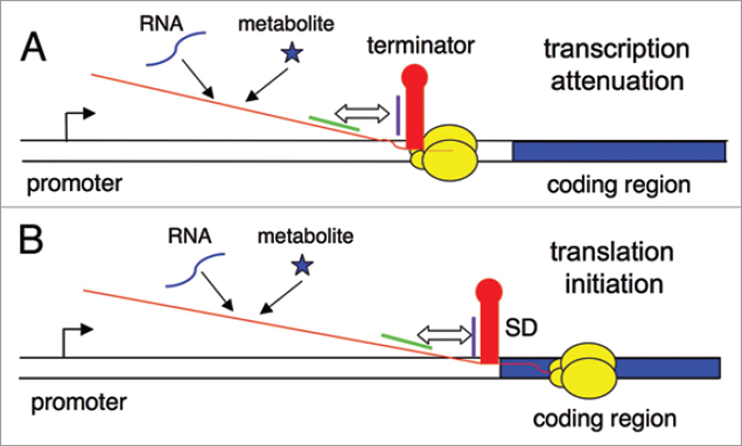
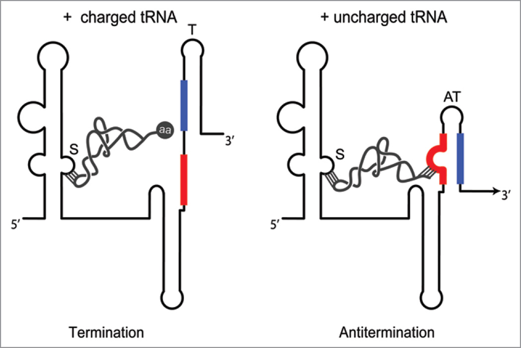
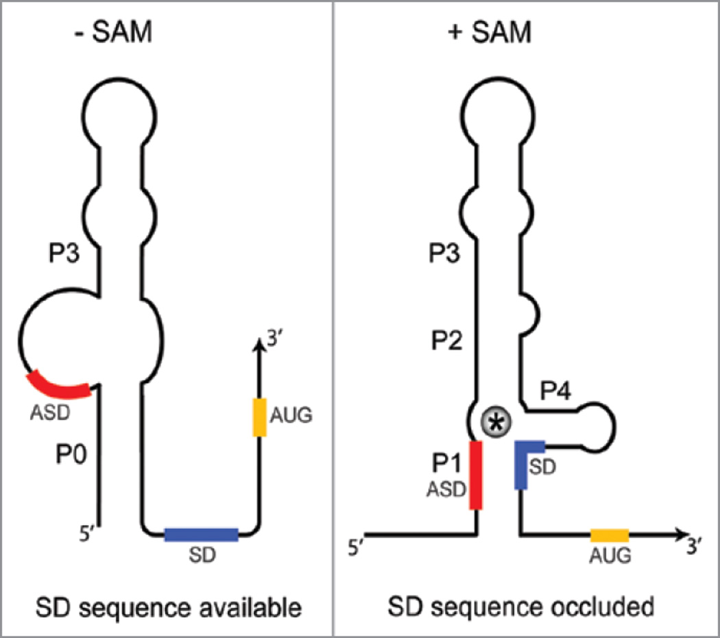
Riboswitches are cis-encoded, cis-acting RNA elements that directly sense a physiological signal. Signal response results in a change in RNA structure that impacts gene expression. Elements of this type play an important role in bacteria, where they regulate a variety of fundamental cellular pathways. Riboswitch-mediated gene regulation most commonly occurs by effects on transcription attenuation, to control whether a full-length transcript is synthesized, or on translation initiation, in which case the transcript is constitutively synthesized but binding of the translation initiation complex is modulated. An overview of the role of riboswitch RNAs in bacterial gene expression will be provided, and a few examples are described in more detail to illustrate the types of mechanisms that have been uncovered.


Figures

Figure 1
Figure 1
The riboswitch mechanism. DNA is shown as a double line, the promoter region is shown as a bent arrow, the regulated coding sequence is shown as a blue rectangle, RNAP is shown as yellow ovals, and the nascent transcript is a red line. (A) Regulation of transcription attenuation. Genes that are regulated at this level include an intrinsic terminator (red stem-loop) in the leader region, upstream of the regulated coding sequence. If this helix forms in the nascent RNA when RNAP is paused during synthesis of the U run (dotted red line), RNAP will terminate transcription. Pairing of sequences on the 5' side of the terminator (purple line) with complementary sequences that are further upstream (green line) results in formation of an alternate antiterminator structure. Binding of regulatory factors (RNA or metabolites) determines whether the RNA folds into the terminator or antiterminator structure. (B) Regulation of translation initiation. Genes that are regulated at this level include a structure (red stem-loop) that can sequester the SD sequence, which results in inhibition of binding of the 30S ribosomal subunit. Pairing of anti-SD (ASD) sequences (purple line) with complementary sequences that are further upstream (green line) results in formation of an alternate structure that sequesters the ASD sequence. Binding of regulatory factors (RNA or metabolites) determines whether the RNA folds into the ASD-SD structure or the competing structure that sequesters the ASD sequence.
Figure 2
Figure 2
The T box riboswitch. Regulation at the level of transcription attenuation, which is the most common mechanism, is shown. The riboswitch senses the relative amounts of uncharged and charged tRNA, for a specific tRNA species, which is recognized primarily by codon-anticodon pairing at the Specifier Sequence (S). Charged tRNA (left) can interact only at the Specifier Sequence; the presence of the amino acid (aa) at the 3' end of the tRNA blocks pairing of the acceptor end of the tRNA with the antiterminator bulge; the terminator helix (T, blue-black) forms, and transcription terminates before RNAP transcribes the downstream coding sequence. Uncharged tRNA (right) can interact at both the Specifier Sequence and the antiterminator (AT) bulge; pairing at the antiterminator stabilizes that structure (red-blue), which sequesters sequences (blue) that would otherwise participate in formation of the terminator helix. Transcription continues (arrow), and the downstream coding sequence is transcribed. In genes regulated at the level of translation initiation, transcription is predicted to be constitutive and the terminator helix is replaced by an ASD-SD helix that sequesters the SD sequence and prevents binding of the ribosome. tRNA binding stabilizes a competing structure analogous to the antiterminator that sequesters the ASD sequence, and frees the SD sequence to allow access of the ribosome.
Figure 3
Figure 3
The S box riboswitch. Regulation at the level of transcription attenuation, which is the more common mechanism, is shown. The SAM-binding aptamer is comprised of helices P1–P4. When SAM levels are low, the more stable antiterminator structure (AT, red-blue) forms, and transcription continues into the downstream coding sequence (arrow head). When SAM levels are high, SAM (*) binds in a pocket formed by stacking of the P1 and P3 helices; this stabilizes the P1 helix, which serves as an anti-antiterminator (AAT, black-red) because it sequesters sequences (red) that would otherwise participate in formation of the antiterminator. Inhibition of antiterminator formation releases sequences from the 3' side of the antiterminator (blue) for formation of the terminator helix (blue-black), and transcription terminates. The SAM binding pocket is stabilized by tertiary interactions including the pseudoknot formed between the loop of P2 and the junction between helices P3 and P4 (dashed line). In genes regulated at the level of translation initiation, transcription is predicted to be constitutive and the terminator helix is replaced by an ASD-SD helix that sequesters the SD sequence and prevents binding of the ribosome. SAM binding stabilizes a structure analogous to the anti-antiterminator that sequesters sequences that would otherwise pair with the ASD sequence, and frees the ASD sequence to pair with the SD to block access of the ribosome.
Figure 4
Figure 4
The SMK box riboswitch. Regulation is at the level of translation initiation for all SMK box riboswitches uncovered to date. In the absence of SAM, the SD sequence is available for binding of the 30S ribosomal subunit, and sequences at the 5' end pair to form the P0 helix. Binding of SAM (*) results in disruption of the P0 helix and sequestration of the SD sequence in the ASD-SD helix (P1). SAM makes direct contacts with residues within the SD. Sequestration of the SD sequence prevents ribosome binding, resulting in repression of gene expression.

Similar articles

Cited by

References

    1. Grundy FJ, Henkin TM. From ribosome to riboswitch: control of gene expression in bacteria by RNA structural rearrangements. Crit Rev Biochem Mol Biol. 2006;41:329–338. - PubMed
    1. Henkin TM. Riboswitch RNAs: using RNA to sense cellular metabolism. Genes Dev. 2008;22:3383–3390. - PMC - PubMed
    1. Montange RK, Batey RT. Riboswitches: emerging themes in RNA structure and function. Annu Rev Biophys. 2008;37:117–133. - PubMed
    1. Dambach MD, Winkler WC. Expanding roles for metabolite-sensing regulatory RNAs. Curr Opin Microbiol. 2009;12:161–169. - PMC - PubMed
    1. Storz G, Opdyke JA, Wassarman KM. Regulating bacterial transcription with small RNAs. Cold Spring Harbor Symp Quant Biol. 2006;71:269–273. - PubMed

Publication types

MeSH terms

Substances

LinkOut - more resources