. 2009 Mar-Apr;1(2):128-41.
doi: 10.4161/mabs.1.2.7631. Epub 2009 Mar 11.

Anti-tumor activity of stability-engineered IgG-like bispecific antibodies targeting TRAIL-R2 and LTbetaR

Affiliations

Anti-tumor activity of stability-engineered IgG-like bispecific antibodies targeting TRAIL-R2 and LTbetaR

Jennifer S Michaelson et al. MAbs. 2009 Mar-Apr.

Abstract

Bispecific antibodies (BsAbs) represent an emerging class of biologics that achieve dual targeting with a single agent. Recombinant DNA technologies have facilitated a variety of creative bispecific designs with many promising therapeutic applications; however, practical methods for producing high quality BsAbs that have good product stability, long serum half-life, straightforward purification, and scalable production have largely been limiting. Here we describe a protein-engineering approach for producing stable, scalable tetravalent IgG-like BsAbs. The stability-engineered IgG-like BsAb was envisioned to target and crosslink two TNF family member receptors, TRAIL-R2 (TNF-Related Apoptosis Inducing Ligand Receptor-2) and LTbetaR (Lymphotoxin-beta Receptor), expressed on the surface of epithelial tumor cells with the goal of triggering an enhanced anti-tumor effect. Our IgG-like BsAbs consists of a stability-engineered anti-LTbetaR single chain Fv (scFv) genetically fused to either the N- or C-terminus of the heavy chain of a fulllength anti-TRAIL-R2 IgG1 monoclonal antibody. Both N- or C-terminal BsAbs were active in inhibiting tumor cell growth in vitro, and with some cell lines demonstrated enhanced activity relative to the combination of parental Abs. Pharmacokinetic studies in mice revealed long serum half-lives for the BsAbs. In murine tumor xenograft models, therapeutic treatment with the BsAbs resulted in reduction in tumor volume either comparable to or greater than the combination of parental antibodies, indicating that simultaneously targeting and cross-linking receptor pairs is an effective strategy for treating tumor cells. These studies support that stability-engineering is an enabling step for producing scalable IgG-like BsAbs with properties desirable for biopharmaceutical development.


Figures

Figure 1
Figure 1
Design and production of IgG-like BsAbs. (A and B), Schematic diagrams of N- and C-BsAbs designs and mammalian expression vectors used for producing IgG-like BsAbs. Detailed components of the expression vectors are shown at the bottom of (B). (C), Analytical size-exclusion chromatography profile of C-BsAb constructed with wild-type BHA10 scFv following expression in CHO cells and purification on Protein A.
Figure 2
Figure 2
DSC analysis of BHA10 FAb and wild-type BHA10 scFv. Overlay of BHA10 FAb (dashed line) and wild-type scFv (solid line) thermograms. Unfolding transitions are measured at the endothermic peaks (transition midpoints) and reported as Tm values (°C).
Figure 3
Figure 3
Molecular model and sequence of BHA10 scFv. (A) The scFv is in the VH□VL orientation. VH is shown in red, VL is shown in blue, and the Gly/Ser linker is shown in red. Paired cysteine substitutions are shown at VH and VL positions 44 and 100, and 105 and 43, respectively. The VH 105C is shown unpaired as it utilizes VH 44C. (B) Amino acid sequence of wild-type BHA10 scFv. The Gene III signal peptide is shown underlined, (Gly4Ser)3 linker is indicated in bold type, and the Enterokinase site, Myc and His tags are indicated in italics. Positions of cysteine substitutions are as follows—the VH substitution at position 44 is shown as a single underline and at position 105 as a double underline. The VL substitution at position 43 is shown as a single underline and at positions 100 and 105 as double underlines.
Figure 4
Figure 4
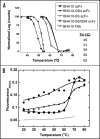
Characterization of stability-engineered BHA10 scFvs. (A) Binding activity of wild-type BHA10 scFv, stability-engineered BHA10 scFvs, and BHA10 FAb to LTβR was measured following thermal challenge. Binding profiles are normalized to 100% maximum binding. The temperature at which 50% binding activity is retained (T50, °C) is indicated. Samples were assayed in duplicate. (B) Temperature-dependent binding of the hydrophobic dye ANS to BHA10 scFvs. PBS, (○); wild-type BHA10 scFv (containing (Gly4Ser)3 linker), (●); BHA10-GS4 scFv (▴); and BHA10-SS/GS4 scFv (▪).
Figure 5
Figure 5
Design and expression of stability-engineered BsAbs. (A) Western blot analysis of BsAbs transiently expressed in CHO cells. Supernatant samples in the left panel are analyzed under non-reducing conditions and under reducing conditions in the right panel. Lane 1: MW marker, 2: C-BsAb, 3: C-BsAb-GS4, 4: C-BsAb-SS, 5: C-BsAb-SS/GS4, 6: N-BsAb, 7: N-BsAb-GS4, 8: N-BsAb-SS, 9: N-BsAb-SS/GS4. Arrow indicates presence of ∼55–60 kDa unidentified immunoreactive species. (B) Normalized SEC profiles of stability-engineered C-BsAbs following Protein A chromatography. Sharp peaks eluting between 13–15 min represent monomeric BsAb. Broad peaks eluting between ∼10.75–14 min represent aggregated protein. Blue dash-dot = C-BsAb, red dash = C-BsAb-SS, and green line = C-BsAb-SS/GS4.
Figure 6
Figure 6
Intact mass analyses of N- and C-BsAb-SS/GS4. N- and C-BsAb-SS/GS4 stored for T = 0 and T = 3 months at 4°C were analyzed for proteolysis and post-translational modification by LC/MS. Upper, N-BsAb-SS/GS light and heavy chains; lower, C-BsAb-SS/GS light and heavy chains. Both light chain and heavy chain masses are consistent with theoretical calculations based upon protein primary sequences. No significant changes in mass as results of modifications were detected. Typical IgG biantennary glycans with variable numbers of terminal galactose were observed such as G0, G1 and G2.
Figure 7
Figure 7
Dual-binding activity of stability-engineered BsAbs. Surface plasmon resonance analysis of stability-engineered BsAbs for dual-binding to TRAIL-R2 and LTβR. Biotinylated anti-His6 antibody was immobilized onto a streptavidin-labeled sensorchip followed by capture of TRAIL-R2-Fc-His6 (TRAIL-R2). (A) sensorgrams show subsequent additions of BsAbs and 0, 3, 10, 30 or 100 nM concentrations of sLTβR-Fc (LTβR). N-BsAb-SS/GS4 shown as dashed line and C-BsAb-SS/GS4 shown as solid line. (B) calculated kinetic rate constants.
Figure 8
Figure 8
Inhibition of tumor cell line proliferation by stability-engineered BsAbs. Viability of human tumor cell lines and control HUVEC cells in response to treatment with BsAb and parent mAbs in a 96-well proliferation assay. (A) WiDr colon tumor cell line; (B) MDA-MB231 breast tumor cell line; (C) Me180 breast tumor cell line; and (D) HUVEC normal umbilical vein endothelial cells. Data is representative of triplicate experiments and shown as mean ± S.D.
Figure 9
Figure 9
Binding of BsAbs to neonatal Fc receptor. Binding of various concentrations of biotinylated FcRn-Fc to fixed concentrations of immobilized N-BsAb-SS/GS4 BsAb (+), N-BSAb-SS/GS4 BsAb (□), hIgG1 (◆) and mIgG1 (▴) in an ELISA assay. Receptor complexes were washed with PBS, pH 6, detected by incubating with streptavidin-HRP conjugate and developed with peroxidase substrate. Plates were read at 450 nm. (A) mouse FcRn-Fc; and (B) human FcRn-Fc.
Figure 10
Figure 10
Inhibition of tumor growth by stability-engineered BsAbs. (A) WiDr and (B) MDA-MB231 tumor cells were implanted subcutaneously into immunocompromised mice. Treatment was initiated on day 13 when tumor size reached approximately 100 mm3. Vehicle control and individual or mixtures of mAbs BHA10 and 14A2 were administered by ip injection 2×/week throughout the study. BsAbs were administered by ip injection 1 ×/week throughout the study. Tumor growth curves are each derived from cohorts consisting of ten mice. Data is represented as mean tumor volume ± S.D.

Similar articles

Cited by

References

    1. Fischer N, Leger O. Bispecific antibodies: molecules that enable novel therapeutic strategies. Pathobiology. 2007;74:3–14. - PubMed
    1. Boerman OC, van Schaijk FG, Oyen WJ, Corstens FH. Pretargeted radioimmunotherapy of cancer: progress step by step. J Nucl Med. 2003;44:400–411. - PubMed
    1. Jain M, Kamal N, Batra SK. Engineering antibodies for clinical applications. Trends Biotechnol. 2007;25:307–316. - PubMed
    1. Kontermann RE. Recombinant bispecific antibodies for cancer therapy. Acta Pharmacol Sin. 2005;26:1–9. - PubMed
    1. Marvin JS, Zhu Z. Recombinant approaches to IgG-like bispecific antibodies. Acta Pharmacol Sin. 2005;26:649–658. - PubMed

MeSH terms

Substances

LinkOut - more resources