

High level expression of functional human IgMs in human PER.C6 cells
- PMID: 20061826
- PMCID: PMC2725415
- DOI: 10.4161/mabs.1.2.7945
High level expression of functional human IgMs in human PER.C6 cells
Abstract
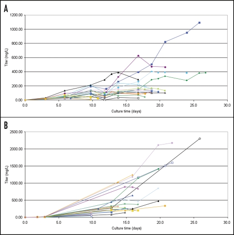
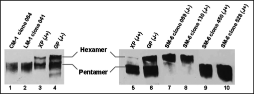
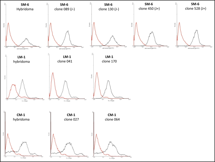
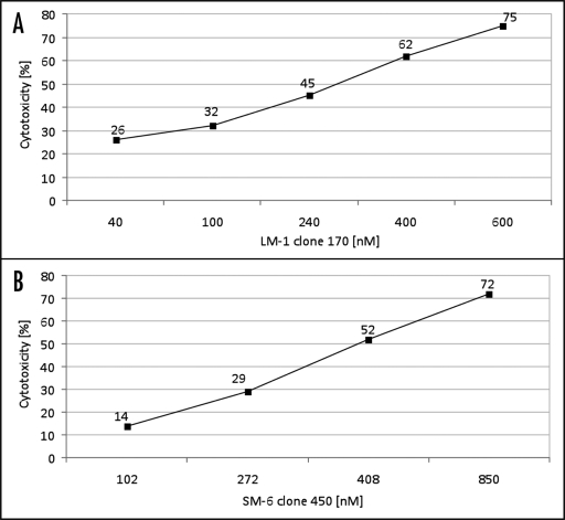
Natural IgM antibodies play an important role in the body's defense mechanisms against transformed cells in the human body and are currently being exploited both in prognoses of malignant lesions and in the therapy of cancer patients. However, despite growing interest and clinical promise, thus far the IgM class of antibodies has failed to gain widespread commercial interest as these are considered to be difficult to produce recombinantly. IgMs are polymeric and have a relatively large mass. In addition, IgM molecules are heavily glycosylated and, when produced in non-human cell lines, they may contain non-human glycan structures which may be potentially immunogenic. Clearly, production systems capable of expressing human recombinant IgM antibodies are needed. We have successfully used PER.C6 cells-a human cell line-to generate three separate human recombinant monoclonal IgMs in suspension cultures in protein-free medium. All three of the IgMs were constructed with joining (J) chain and were expressed in the pentameric form. One of the IgMs was also expressed as a hexamer without J chain. Clones with cell specific productivities greater than 20 pg/cell/day were generated, which led to yields of 0.5 g/L to 2g/L in fed-batch production. All the IgMs expressed were biologically active as shown in binding and cytotoxicity assays. These studies demonstrate the potential of PER.C6 cells for the production of high levels of functional recombinant IgM and other polymeric molecules, using a straightforward and rapid stable cell line generation method.
Figures
Similar articles
-
Generation of stable cell clones expressing mixtures of human antibodies.Biotechnol Bioeng. 2010 Aug 1;106(5):741-50. doi: 10.1002/bit.22763. Biotechnol Bioeng. 2010. PMID: 20564612
-
Recombinant human hexamer-dominant IgM monoclonal antibody to ganglioside GM3 for treatment of melanoma.Clin Cancer Res. 2007 May 1;13(9):2745-50. doi: 10.1158/1078-0432.CCR-06-2919. Clin Cancer Res. 2007. PMID: 17473208
-
Glyco engineered pentameric SARS-CoV-2 IgMs show superior activities compared to IgG1 orthologues.Front Immunol. 2023 Jun 8;14:1147960. doi: 10.3389/fimmu.2023.1147960. eCollection 2023. Front Immunol. 2023. PMID: 37359564 Free PMC article.
-
Structure, Function, and Therapeutic Use of IgM Antibodies.Antibodies (Basel). 2020 Oct 13;9(4):53. doi: 10.3390/antib9040053. Antibodies (Basel). 2020. PMID: 33066119 Free PMC article. Review.
-
Advances in recombinant antibody manufacturing.Appl Microbiol Biotechnol. 2016 Apr;100(8):3451-61. doi: 10.1007/s00253-016-7388-9. Epub 2016 Mar 3. Appl Microbiol Biotechnol. 2016. PMID: 26936774 Free PMC article. Review.
Cited by
-
Comprehensive method for producing high-affinity mouse monoclonal antibodies of various isotypes against (4-hydroxy-3-nitrophenyl)acetyl (NP) hapten.Heliyon. 2024 Nov 29;10(23):e40837. doi: 10.1016/j.heliyon.2024.e40837. eCollection 2024 Dec 15. Heliyon. 2024. PMID: 39698082 Free PMC article.
-
Identification of Novel Epitopes with Agonistic Activity for the Development of Tumor Immunotherapy Targeting TRAIL-R1.J Cancer. 2017 Aug 2;8(13):2542-2553. doi: 10.7150/jca.19918. eCollection 2017. J Cancer. 2017. PMID: 28900492 Free PMC article.
-
The role of magnetic field in the biopharmaceutical production: Current perspectives.Biotechnol Rep (Amst). 2019 Apr 4;22:e00334. doi: 10.1016/j.btre.2019.e00334. eCollection 2019 Jun. Biotechnol Rep (Amst). 2019. PMID: 31011551 Free PMC article. Review.
-
IgM production by bone marrow plasmablasts contributes to long-term protection against intracellular bacterial infection.J Immunol. 2011 Jan 15;186(2):1011-21. doi: 10.4049/jimmunol.1002836. Epub 2010 Dec 8. J Immunol. 2011. PMID: 21148037 Free PMC article.
-
Transient pentameric IgM fulfill biological function-Effect of expression host and transfection on IgM properties.PLoS One. 2020 Mar 12;15(3):e0229992. doi: 10.1371/journal.pone.0229992. eCollection 2020. PLoS One. 2020. PMID: 32163462 Free PMC article.
References
-
- Kohler G, Milstein C. Continuous cultures of fused cells secreting antibody of predefined specificity. Nature. 1975;256:495–497. - PubMed
-
- Reichert JM, Valge-Archer VE. Development trends for monoclonal antibody cancer therapeutics. Nature Reviews. 2007;6:349–356. - PubMed
-
- Collins C, Tsui FWL, Shulman MJ. Differential activation of human and guinea pig complement by pentameric and hexameric IgM. Eur J Immunol. 2002;32:1802–1810. - PubMed
-
- Wiersma EJ, Collins C, Fazel S, Shulman MJ. Structural and functional analysis of J chain-deficient IgM. J Immunol. 1998;160:5979–5989. - PubMed
MeSH terms
Substances
LinkOut - more resources
Full Text Sources
Other Literature Sources