. 2010 Feb;13(2):239-45.
doi: 10.1038/nn.2475. Epub 2010 Jan 10.

A neural mechanism for exacerbation of headache by light

Affiliations

A neural mechanism for exacerbation of headache by light

Rodrigo Noseda et al. Nat Neurosci. 2010 Feb.

Abstract

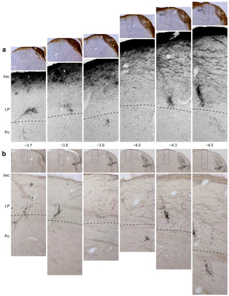
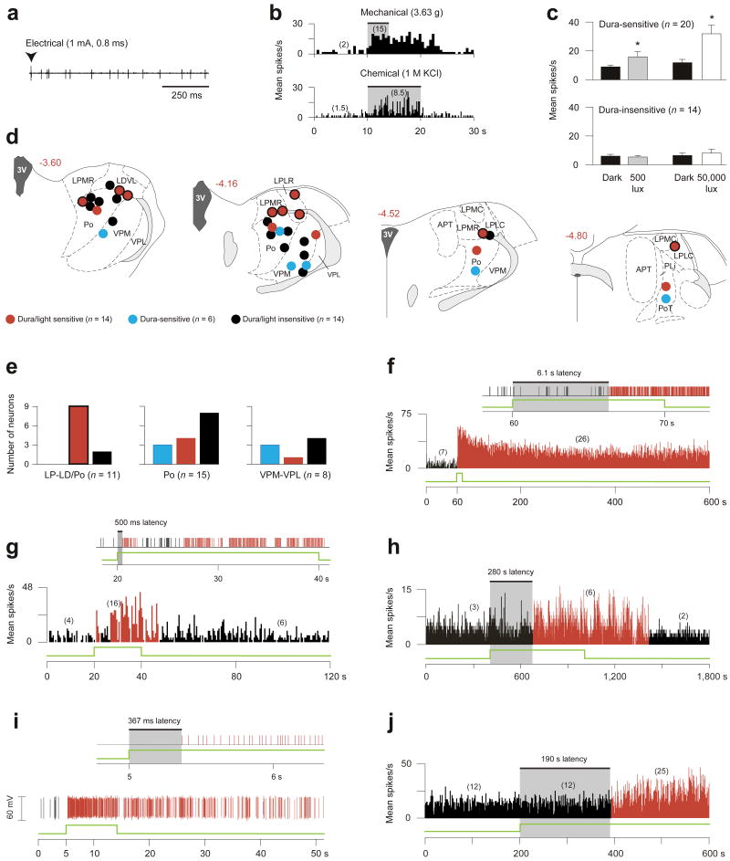
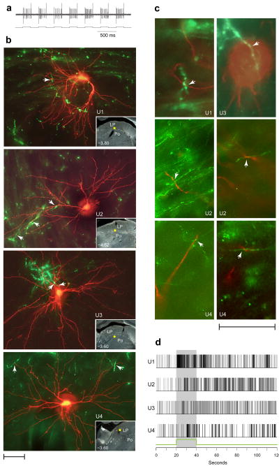
The perception of migraine headache, which is mediated by nociceptive signals transmitted from the cranial dura mater to the brain, is uniquely exacerbated by exposure to light. We found that exacerbation of migraine headache by light is prevalent among blind individuals who maintain non-image-forming photoregulation in the face of massive rod/cone degeneration. Using single-unit recording and neural tract tracing in the rat, we identified dura-sensitive neurons in the posterior thalamus whose activity was distinctly modulated by light and whose axons projected extensively across layers I-V of somatosensory, visual and associative cortices. The cell bodies and dendrites of such dura/light-sensitive neurons were apposed by axons originating from retinal ganglion cells (RGCs), predominantly from intrinsically photosensitive RGCs, the principle conduit of non-image-forming photoregulation. We propose that photoregulation of migraine headache is exerted by a non-image-forming retinal pathway that modulates the activity of dura-sensitive thalamocortical neurons.


Figures

Figure 1
Figure 1
Projections of retinal ganglion cells to the lateral posterior thalamic nuclei (LP) and posterior thalamic nuclear group (Po). Retrograde tracing of retinal afferents was performed using large (a) or small (b) injections of rAAV–GFP into the vitreous body of the eye (see text for details). (a) Top panels: low-power images of coronal sections counterstained with thionin, showing immunolabeled retinal afferents (brown) in the main visual pathway. Bottom panels: high-power detail of the boxed areas in the corresponding top panels, showing retinal afferents in ventral LP and dorsal Po. These images display only the blue channel which isolated the labeled fibers from the background Nissl staining. (b) Top panels: low-power images of coronal sections showing preferential labeling of retinal afferents in the intergeniculate leaflet (IGL). Bottom panels: high-power detail of the boxed areas in the corresponding top panels, showing immunolabeled retinal afferents in ventral LP and dorsal Po. Preferential labeling of non-image-forming pathways by the smaller rAAV–GFP injection (b) compared with the larger injection (a) reflects preferential labeling of ipRGC (see text for explanation). Note that the retinal afferents run dorsoventrally from the main visual pathway through ventral aspect of LP and into dorsal aspect of Po (a,b). Note that the density of labeled axons in ventral LP and dorsal Po is similar between a and b, suggesting that most labeled axons in these areas were of ipRGC origin. Numbers indicate distance from Bregma (mm). Abbreviations: APT, anterior pretectal nucleus; bsc, brachium superior colliculus; DLG, dorsal part of lateral geniculate nucleus. Scale bars represent 500 μm.
Figure 2
Figure 2
Photosensitivity of dura-sensitive thalamic neurons. (a,b) Identifying neuronal responses to electrical (a), mechanical and chemical (b) stimulation of the dura. (c) Effects of ambient light (500 lux) and bright light (50,000 lux) on firing rate (mean ± s.e.m.) of dura-sensitive vs. dura-insensitive thalamic neurons (*P < 0.05; Wilcoxon matched-pairs signed-ranks test). (d) Histological localization of the recorded neurons. Drawings and numbers indicating distance from Bregma (mm) are based on Paxinos and Watson. For abbreviations see text. (e) Graphic representation of the dorso-ventral localization of the neurons shown in d. Color coding same as in d; bordered red bar in left panel corresponds to bordered red circles in d. (f–j) Examples of delayed and immediate photoactivation of individual dura-sensitive thalamic neurons by 50,000 (f,g), 3,000 (h,i) and 500 lux (j) of white light (green line). Panels f, g consist of window discriminator spike output (top) and mean activity histogram (bottom). Panels h, j show mean activity histogram. Panel i consists of window discriminator output (top) and oscillographic tracing (bottom). Each of the light intensities induced delayed activation in some neurons (f,h,j) and immediate activation in others (g,i). Each of the light intensities induced prolonged activation that outlasted the stimulus by several minutes. Numbers in parentheses indicate mean spikes/s for the corresponding interval. Black and red bars indicate, respectively, baseline and enhanced periods of activity in response to light. Bin width are 0.5 (f,g,j) and 1 s (h).
Figure 3
Figure 3
Close apposition between dura/light-sensitive neurons and retinal afferents in LP and Po. (a) Synchronization of neuronal activity (top trace) with the current (bottom trace) delivered by the TMR–dextran filled recording micropipette (see text for detail). (b) Dura/light sensitive units (U1–U4) filled with TMR–dextran (red) and retinal axons labeled anterogradely with CTB (green). Each image represents z-stacking of approximately thirty 1–1.5-μm-thick scans. Arrowheads point to potential axodendritic or axosomatic apposition. Localization of each cell body is marked by a yellow star in the low-power, darkfield inset. Numbers indicate distance from Bregma. (c) Evidence for axodendritic and axosomatic apposition within a single 1–1.5-μm-thick scan taken from the units shown in b. (d) Neuronal firing in response to 50,000 lux of white light (green line and shaded area), corresponding to the individual neurons shown in b. Scale bars represent 50 μm (b,c).
Figure 4
Figure 4
Cortical projections of three dura/light-sensitive thalamic neurons juxtacellularly-filled with TMR–dextran. (a) Labeled cell bodies and their dendrites in the posterior thalamus. (b) Camera-lucida tracing of the cell bodies, dendrites, and axonal trajectories coursing through thalamic reticular nucleus (Rt) en route the external capsule (ec). (c) Localization of cell bodies, Rt collaterals and entry point of the parent axon into the external capsule. (d) Tabulation of cortical areas and layers containing axons with synaptic boutons. (e) Camera-lucida tracing of axon terminal fields in different cortical areas. (f) Photomicrographs of axons with synaptic boutons in several cortical areas. Drawings and numbers (b,c,e) indicating distance from Bregma (mm) are based on Paxinos and Watson. Au1, primary auditory cortex; AuD, secondary auditory cortex, dorsal; M1 and M2, primary and secondary motor cortices, respectively; S1, primary somatosensory cortex. For other abbreviations not listed here see text. Scale bars represent 100 μm (a,f).

Comment in

  • Shining a spotlight on headaches.
    Molnár Z, Taylor JS. Molnár Z, et al. Nat Neurosci. 2010 Feb;13(2):150-1. doi: 10.1038/nn0210-150. Nat Neurosci. 2010. PMID: 20104207 No abstract available.

Similar articles

Cited by

References

    1. The International Classification of Headache Disorders, Second Edition. Cephalalgia. 2004;24:1–160. - PubMed
    1. Selby G, Lance JW. Observations on 500 cases of migraine and allied vascular headache. J Neurol Neurosurg Psychiat. 1960:23–32. - PMC - PubMed
    1. Markowitz S, Saito K, Moskowitz MA. Neurogenically mediated plasma extravasation in dura mater: effect of ergot alkaloids. A possible mechanism of action in vascular headache. Cephalalgia. 1988;8:83–91. - PubMed
    1. Penfield W, McNaughton F. Dural headache and innervation of the dura mater. Arch, Neurol Psychiat. 1940;44:43–75.
    1. Burstein R, Yamamura H, Malick A, Strassman AM. Chemical stimulation of the intracranial dura induces enhanced responses to facial stimulation in brain stem trigeminal neurons. Journal of Neurophysiology. 1998;79:964–982. - PubMed

Publication types

MeSH terms