

Design and validation of a clinical instrument for spectral diagnosis of cutaneous malignancy
- PMID: 20062500
- PMCID: PMC2812816
- DOI: 10.1364/AO.49.000142
Design and validation of a clinical instrument for spectral diagnosis of cutaneous malignancy
Abstract
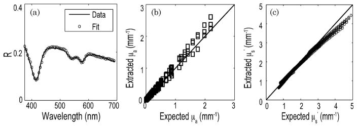
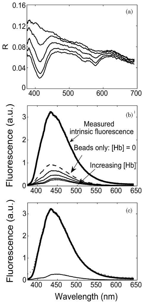
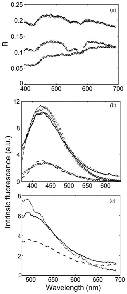
We report a probe-based portable and clinically compatible instrument for the spectral diagnosis of melanoma and nonmelanoma skin cancers. The instrument combines two modalities--diffuse reflectance and intrinsic fluorescence spectroscopy--to provide complementary information regarding tissue morphology, function, and biochemical composition. The instrument provides a good signal-to-noise ratio for the collected reflectance and laser-induced fluorescence spectra. Validation experiments on tissue phantoms over a physiologically relevant range of albedos (0.35-0.99) demonstrate an accuracy of close to 10% in determining scattering, absorption and fluorescence characteristics. We also demonstrate the ability of our instrument to collect in vivo diffuse reflectance and fluorescence measurements from clinically normal skin, dysplastic nevus, and malignant nonmelanoma skin cancer.
Figures
Similar articles
-
Pilot clinical study for quantitative spectral diagnosis of non-melanoma skin cancer.Lasers Surg Med. 2010 Dec;42(10):716-27. doi: 10.1002/lsm.21009. Lasers Surg Med. 2010. PMID: 21246575 Free PMC article. Clinical Trial.
-
A hyperspectral fluorescence lifetime probe for skin cancer diagnosis.Rev Sci Instrum. 2007 Dec;78(12):123101. doi: 10.1063/1.2818785. Rev Sci Instrum. 2007. PMID: 18163714
-
In-vivo characterization of optical properties of pigmented skin lesions including melanoma using oblique incidence diffuse reflectance spectrometry.J Biomed Opt. 2011 Feb;16(2):020501. doi: 10.1117/1.3536509. J Biomed Opt. 2011. PMID: 21361657 Free PMC article.
-
Spectroscopic methods for the photodiagnosis of nonmelanoma skin cancer.J Biomed Opt. 2013 Jun;18(6):061221. doi: 10.1117/1.JBO.18.6.061221. J Biomed Opt. 2013. PMID: 23748702 Review.
-
Computer-assisted diagnosis techniques (dermoscopy and spectroscopy-based) for diagnosing skin cancer in adults.Cochrane Database Syst Rev. 2018 Dec 4;12(12):CD013186. doi: 10.1002/14651858.CD013186. Cochrane Database Syst Rev. 2018. PMID: 30521691 Free PMC article.
Cited by
-
Properties of contact pressure induced by manually operated fiber-optic probes.J Biomed Opt. 2015;20(12):127002. doi: 10.1117/1.JBO.20.12.127002. J Biomed Opt. 2015. PMID: 26720880
-
Sampling depth of a diffuse reflectance spectroscopy probe for in-vivo physiological quantification of murine subcutaneous tumor allografts.J Biomed Opt. 2018 Aug;23(8):1-14. doi: 10.1117/1.JBO.23.8.085006. J Biomed Opt. 2018. PMID: 30152204 Free PMC article.
-
Measurements of extrinsic fluorescence in Intralipid and polystyrene microspheres.Biomed Opt Express. 2014 Jul 22;5(8):2726-35. doi: 10.1364/BOE.5.002726. eCollection 2014 Aug 1. Biomed Opt Express. 2014. PMID: 25136497 Free PMC article.
-
Broadband absorption and reduced scattering spectra of in-vivo skin can be noninvasively determined using δ-P1 approximation based spectral analysis.Biomed Opt Express. 2015 Jan 9;6(2):443-56. doi: 10.1364/BOE.6.000443. eCollection 2015 Feb 1. Biomed Opt Express. 2015. PMID: 25780735 Free PMC article.
-
Multimodal Imaging and Spectroscopy Fiber-bundle Microendoscopy Platform for Non-invasive, In Vivo Tissue Analysis.J Vis Exp. 2016 Oct 17;(116):54564. doi: 10.3791/54564. J Vis Exp. 2016. PMID: 27805585 Free PMC article.
References
-
- American Cancer Society. Cancer facts and figures 2009. http://www.cancer.org/docroot/STT/content/STT_1x_Cancer_Facts_Figures_20....
-
- Mogensen M, Jemec G. Diagnosis of nonmelanoma skin cancer/keratinocyte carcinoma: a review of diagnostic accuracy of nonmelanoma skin cancer diagnostic tests and technologies. Dermatol Surg. 2007;33:1158–1174. - PubMed
-
- Zonios G, Perelman LT, Backman V, Manoharan R, Fitzmaurice M, Van Dam J, Feld MS. Diffuse reflectance spectroscopy of human adenomatous colon polyps in vivo. Appl Opt. 1999;38:6628–6637. - PubMed
-
- Marchesini R, Cascinelli N, Brambilla M, Clemente C, Mascheroni L, Pignoli E, Testori A, Venturoli D. In vivo spectrophotometric evaluation of neoplastic and non-neoplastic skin pigmented lesions. II: Discriminant analysis between nevus and melanoma. Photochem Photobiol. 1992;55:515–522. - PubMed
-
- Mourant JR, Bigio IJ, Boyer J, Conn RL, Johnson T, Shimada T. Spectroscopic diagnosis of bladder cancer with elastic light scattering. Lasers Surg Med. 1995;17:350–357. - PubMed
Publication types
MeSH terms
Grants and funding
LinkOut - more resources
Full Text Sources
Medical
Molecular Biology Databases
