. 2010 Jan;6(1):e1000804.
doi: 10.1371/journal.pgen.1000804. Epub 2010 Jan 8.

VEZF1 elements mediate protection from DNA methylation

Affiliations

VEZF1 elements mediate protection from DNA methylation

Jacqueline Dickson et al. PLoS Genet. 2010 Jan.

Abstract

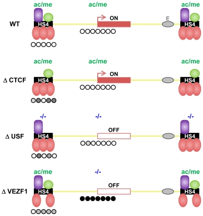
There is growing consensus that genome organization and long-range gene regulation involves partitioning of the genome into domains of distinct epigenetic chromatin states. Chromatin insulator or barrier elements are key components of these processes as they can establish boundaries between chromatin states. The ability of elements such as the paradigm beta-globin HS4 insulator to block the range of enhancers or the spread of repressive histone modifications is well established. Here we have addressed the hypothesis that a barrier element in vertebrates should be capable of defending a gene from silencing by DNA methylation. Using an established stable reporter gene system, we find that HS4 acts specifically to protect a gene promoter from de novo DNA methylation. Notably, protection from methylation can occur in the absence of histone acetylation or transcription. There is a division of labor at HS4; the sequences that mediate protection from methylation are separable from those that mediate CTCF-dependent enhancer blocking and USF-dependent histone modification recruitment. The zinc finger protein VEZF1 was purified as the factor that specifically interacts with the methylation protection elements. VEZF1 is a candidate CpG island protection factor as the G-rich sequences bound by VEZF1 are frequently found at CpG island promoters. Indeed, we show that VEZF1 elements are sufficient to mediate demethylation and protection of the APRT CpG island promoter from DNA methylation. We propose that many barrier elements in vertebrates will prevent DNA methylation in addition to blocking the propagation of repressive histone modifications, as either process is sufficient to direct the establishment of an epigenetically stable silent chromatin state.


Conflict of interest statement

The authors have declared that no competing interests exist.

Figures

Figure 1
Figure 1. Schematic representation of the chicken β-globin cluster and surrounding loci.
Boxes represent the folate receptor (FR), β-globin (ρ, βH, βA and ε) and chicken olfactory receptor (COR) genes (not to scale). Arrows indicate DNaseI hypersensitive sites. The core of the HS4 element is expanded to show the positions of the five in vitro footprinted sequences .
Figure 2
Figure 2. Three footprinted sites in the HS4 barrier protect a promoter from DNA methylation.
(A) Schematic representation of the IL-2R transgene drawn to scale with the distribution of CpG dinucleotides shown below. Promoter region CpG dinucleotides subject to bisulfite genomic sequencing (BSEQ) analysis are indicated by the gray bar. (B) CpG methylation of transgene promoters flanked by wild-type or mutant HS4 insulators after 30 (left) or 90 (right) days of culture. The percentage of methylation for each CpG from 10 clones is plotted. The average methylation for all CpGs is indicated to the right of each plot. Scoring from individual clones can be found in Table S1. Numbers below each histogram refer to CpG numbering from , where CpG 4–11 and 12–18 reside in the promoter and coding sequence, respectively. Data are representative of two independent transgenic lines, with an average methylation variation of 7% or less. (C) Transgenic IL-2R expression for each line in (B) as monitored by flow cytometry. WT and ΔII lines retain IL-2R expression, whereas ΔI, ΔIII, ΔIV and ΔV all succumb to silencing following 20–40 days of culture.
Figure 3
Figure 3. Mutations of insulator protein binding sites result in the de novo methylation of HS4 itself.
(A) Schematic representation of the IL-2R transgene including the upstream HS4 elements subject to bisulfite genomic sequencing analysis (gray horizontal bar below CpG plot). (B) Schematic representation of a 275 bp HS4 core element, drawn to scale. Footprinted sequences are shaded gray. CpG locations with their assigned numbers are shown above. Horizontal bars indicate bases subject to deletion. (C) CpG methylation of wild-type (WT) or mutant (ΔI - ΔV) HS4 insulators after 30 (left) or 90 (right) days of culture. Both copies of HS4 were sequenced, except for ΔII and ΔV, where only the outermost copy was sequenced (see Materials and Methods). The percentage of methylation for each CpG from 10 clones is plotted. The average methylation for all CpGs is indicated to the right of each plot. Scoring from individual clones can be found in Table S2. Numbers below each histogram refer to CpG numbering as assigned in (B). Horizontal gray bars indicate deleted CpGs. Data are representative of at least two independent transgenic lines, with an average methylation variation of 12% or less.
Figure 4
Figure 4. Nuclear proteins specifically interact with the dG-dC strings at HS4 footprints I, III, and V.
(A–C) Gel mobility shift analysis of interactions between chicken adult erythrocyte nuclear extract and 32P-labelled FI (A), FIII (B), and FV (C) oligonucleotide duplexes. Unlabelled competitor duplexes (indicated above each lane) were added at 50 fold molar excess. Arrows indicate footprint sequence-specific complexes. Asterisks indicate a non-specific FI complex in (A) and an FV complex that was not pursued further due to inconsistent abundance in (C). (D) Sequences present in competitor oligonucleotides. Mutations shown in bold lower case. Footprinted bases are indicated by shading. Bases that are essential for maximal binding are underlined; mutations of other bases had no effect on binding (A–C, data not shown).
Figure 5
Figure 5. Purification of FI- and FIII-binding proteins.
Schematic representation of the steps used to purify DNA-binding proteins from adult erythrocyte nuclear protein (NP) extracts that interact with FI and FIII sequences. Numbers in the workflow indicate the salt concentration (mM) at which DNA binding factors eluted from each column. DNA binding activity was tracked using gel mobility shift analysis and the sequence specificity of complex-forming factors was checked by competition analysis following each purification stage (data not shown). FI- and FIII-binding activities exactly co-purified. The eluate pool from the Heparin step was split in two prior to either FI or FIII DNA affinity chromatography. SDS-PAGE of proteins eluted from DNA affinity columns at 400 mM KCl visualized with colloidal Coomassie is shown below. The size of each polypeptide (kDa) is indicated.
Figure 6
Figure 6. VEZF1 specifically interacts with dG–dC strings within HS4 footprints I and III and the βA-globin promoter.
(A) Gel mobility shift analysis of interactions with 32P-labelled HS4 FI, and FIII, or βA-globin promoter ‘glo wt’ oligonucleotide duplexes. Unlabelled competitor duplexes (indicated above each lane) were added at 50 fold molar excess. Adult chicken erythrocyte nuclear extract (ery) or recombinant chicken VEZF1 were used as indicated by brackets above the lanes. Arrows indicate footprint sequence-specific complexes. (B) Gel mobility supershift assays using 32P-labelled FI, FIII, FV, and glo wt oligonucleotide duplexes. Proteins were pre-incubated with anti-VEZF1 antibodies (indicated above each lane) prior to incubation with DNA. VEZF1 supershifts are evidenced by either abrogation of specific complexes (asterisks) and/or formation of low mobility ternary complexes (SS). Antibodies alone do not give rise to complexes with any of the duplexes used (data not shown). Supershift experiments shown are cropped from the full gels shown in Figure S3. (C) Sequences present in oligonucleotides used in gel mobility shift assays. dG–dC strings and their mutations are bold and underlined, respectively.
Figure 7
Figure 7. VEZF1 interacts with the HS4 insulator in vivo.
(A) ChIP analysis of VEZF1 and CTCF interactions at the β-globin locus in (A) 6C2 erythroid progenitor cells or (B) 10 day embryonic erythrocytes. DNA enrichments at the βA promoter or the HS4 and 3′HS insulators were normalized to a negative control located in the 16 kb condensed chromatin region upstream of the β-globin locus.
Figure 8
Figure 8. VEZF1 elements protect a CpG island from de novo DNA methylation.
(A) Scale representation of the 700 bp hamster APRT CpG island (CGI). The positions of AvaII (A) and HpaII (H) restriction sites are indicated. The locations of putative SP1 and VEZF1 binding motifs in the wild type (wt) sequence are shown. The sequence alterations in the mutant (mut) are shown beneath, where all three SP1/VEZF1 motifs are replaced with the VEZF1-specific footprint III (CCCCCCGCATCCCCGA) sequence from the HS4 insulator. (B) ChIP analysis of SP1 and VEZF1 interactions with stably integrated hamster APRT CGI sequences in mouse ES cells. PCR primers flanking either sites 1 and 2 (1&2) or site 3 were used to detect interactions with either the wild type (wt) APRT CGI or the mutant (FIII mut) containing VEZF1-specific sites. PCR products shown are ∼180 bp in size. DNA enrichments relative to input and normalized to no antibody control are shown (C) Methylation specific PCR (MSP) analysis of a stably integrated non-island region of the APRT locus. Genomic DNA was digested with either of the isoschizomers MspI (M) or CpG methylation-sensitive HpaII (H), followed by PCR over the CpG-containing sites. Increased PCR product from HpaII relative to MspI digested DNA demonstrates the presence of de novo methylation at these sites in ES cells. Two representative lines are shown. (D–G) VEZF1 sites are sufficient for the demethylation of the APRT CpG island. Southern blotting of APRT CpG island elements stably integrated into ES cells. Genomic DNA was digested with AvaII alone (A) or with AvaII and HpaII (AH). Digestion of the ∼600 bp AvaII parent fragments with HpaII indicates the absence of DNA methylation. (D,E) Representative lines harboring the wild type APRT CGI that was unmethylated or in vitro methylated with M.SssI prior to integration. (F,G) Two representative lines harboring the mutant APRT CGI, whose SP1 motifs are substituted with VEZF1-specific FIII sites from HS4, that were unmethylated or pre-methylated prior to integration.
Figure 9
Figure 9. Shielding of a transgene by the multi-component HS4 barrier element.
The observed effects of insulator protein binding site mutations on the DNA methylation (this study), histone modification and transcriptional status (this study and [12]) of a transgene insulated by HS4. A schematic representation is shown where HS4 harbors one CTCF (purple), one USF (green) and three VEZF1 (red) binding sites. Transcriptional activity of the reporter gene is indicated with an arrow. The histone modification status is depicted above each transgene, where high and minimal levels of H3ac and H3K4me2 are represented as ac/me and -/-, respectively. The DNA methylation status of the promoter and upstream insulators is indicated below each transgene where open and filled circles represent unmethylated and methylated bases, respectively. Shades of methylation are an approximation of the data presented in Figure 1 and Figure 2. Mutation of the CTCF site disrupts enhancer blocking activity but has no effect on barrier activity of HS4. USF site mutation disrupts the recruitment of active histone modifications, resulting in transcriptional silencing. The promoter remains unmethylated in USF mutants due to the action of VEZF1. VEZF1 site mutations abrogate barrier activity despite USF-mediated recruitment of histone modifications remaining intact. VEZF1 mutants are characterized by complete promoter methylation.

Similar articles

Cited by

References

    1. Kellum R, Schedl P. A position-effect assay for boundaries of higher order chromosomal domains. Cell. 1991;64:941–950. - PubMed
    1. Stalder J, Larsen A, Engel JD, Dolan M, Groudine M, et al. Tissue-specific DNA cleavages in the globin chromatin domain introduced by DNAase I. Cell. 1980;20:451–460. - PubMed
    1. Gaszner M, Felsenfeld G. Insulators: exploiting transcriptional and epigenetic mechanisms. Nat Rev Genet. 2006;7:703–713. - PubMed
    1. Valenzuela L, Kamakaka RT. Chromatin insulators. Annu Rev Genet. 2006;40:107–138. - PubMed
    1. Wallace JA, Felsenfeld G. We gather together: insulators and genome organization. Curr Opin Genet Dev. 2007;17:400–407. - PMC - PubMed

Publication types