

Mucosa associated lymphoid tissue lymphoma of the colon: a case report
- PMID: 20062639
- PMCID: PMC2803979
- DOI: 10.1186/1757-1626-2-9316
Mucosa associated lymphoid tissue lymphoma of the colon: a case report
Abstract
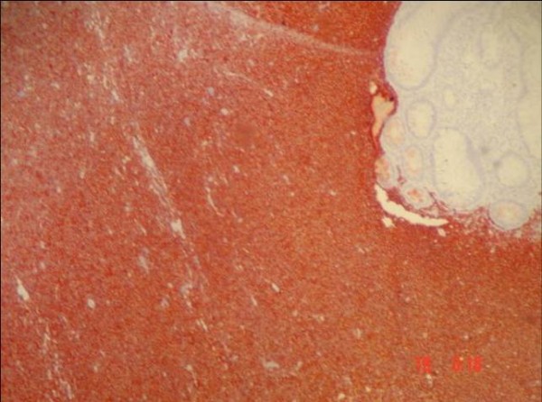
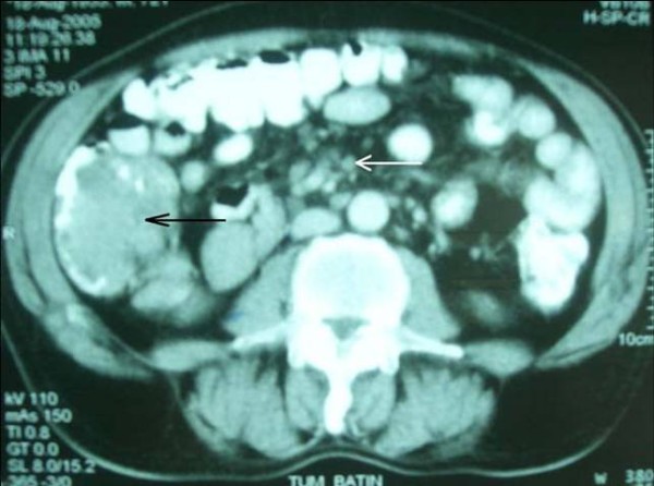
A 65-year-old man had suffered from rectal bleeding during defecation for a few weeks, admitted to our department. Laboratory findings were normal except a slight elevation in the level of alkaline phosphatase. Multiple polypoid lesions were observed in colonoscopic examination. The histological and immunochemical evaluation showed atypical lymphoid cell proliferation and lymphoepithelial lesions on the colonic mucosa, staining with CD20 (CD20 x 100). After the diagnosis had been confirmed as low grade mucosa associated lymphoid tissue lymphoma. Abdominal computed tomography revealed polypoid lesions throughout the colon and multiple milimetrics lymphadenopathies in the mesentery. The patient was treated with a chemotherapy regimen. During the follow-up, colonoscopic examination and blind biopsies were repeated in every 6 months, revealed endoscopically and pathologically normal mucosa each time. The patient is still alive without any recurrence of the disease 36 months after the diagnosis.
Figures


References
-
- Greiner A, Marx A, Heesemann J, Leebmann J, Schmausser B, Muller-Hermelink HK. Idiotype idendity in a MALT-type lyphoma and B-cells in Helcobacter-pyloric associated chronic gastritis. Lab Invest. 1994;70:572–578. - PubMed
-
- Radaszkiewicz T, Dragosics B, Bauer P. Gastrointestinal malignant lymphomas of the mucosa-associated lymphoid tissue: Factors relevant to prognosis. Gastroenterology. 1992;102:1628–1638. - PubMed
LinkOut - more resources
Full Text Sources
