

Radiographic evaluation of bone regeneration after the application of plasma rich in growth factors in a lower third molar socket: a case report
- PMID: 20062651
- PMCID: PMC2803931
- DOI: 10.1186/1757-1626-2-9134
Radiographic evaluation of bone regeneration after the application of plasma rich in growth factors in a lower third molar socket: a case report
Abstract
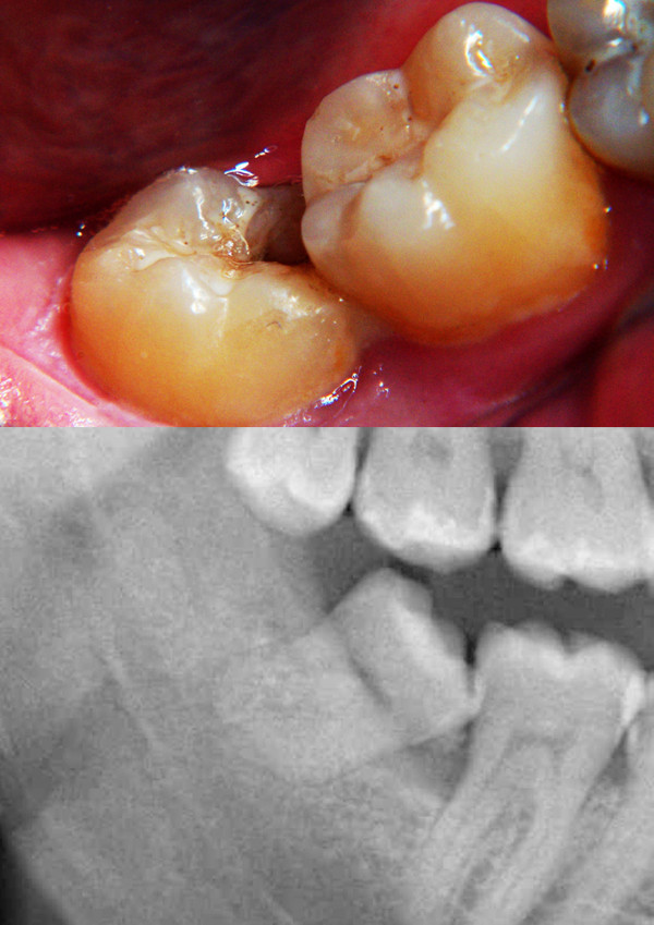
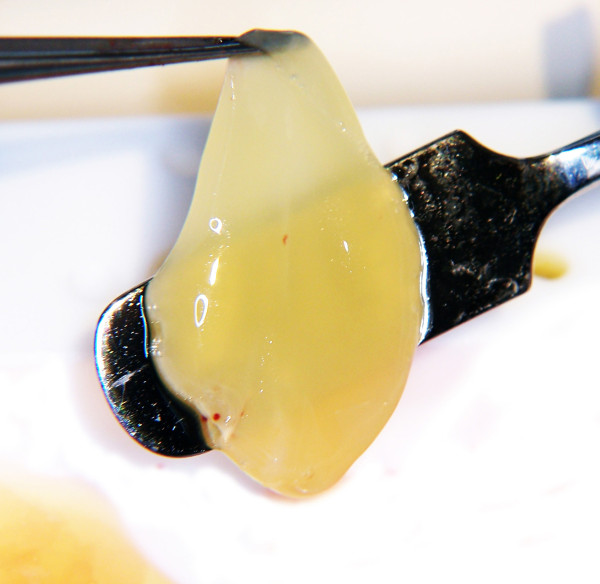
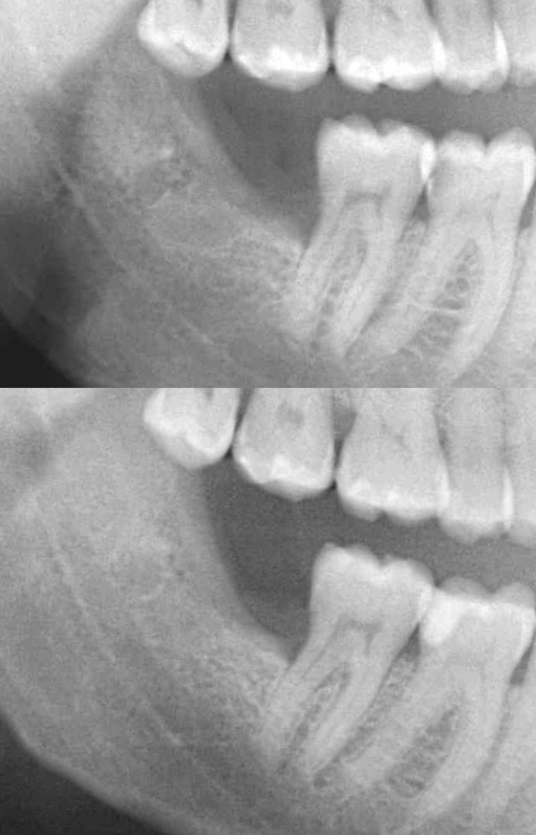
A 42-year-old Mediterranean male presented complaining of inability to sustain good oral care at the posterior aspect of the lower right jaw. The main problems were food impaction in the area and the subsequent malodor. The patient reported remarkable medical history. Clinical examination revealed local erytherma with noticeable bone defect distal to the second molar with obvious defect in the mesial wall of the third molar; the penetration depth was found to be up to 6 mm.Radiological evaluation confirmed the defect and it was attributed to the mesioangularly partially impacted lower third molar. It was decided that third molar should be extracted and concentrate of the patient's growth factors (PRGF) to be applied into the bony defect to stimulate bone regeneration and promote healing.The third molar tooth was, then, removed surgically and the PRGF, which was prepared preoperatively, was implanted in the socket. At the first postoperative day, moderate pain was the main complaint and was controlled by NSAIDs. One week postoperatively, the sutures were removed and there was good tissue healing on examination.On the fiftieth postoperative day, radiographic evaluation took place and showed noticeable enhancement of density and radio-opacity in the third molar socket area, in comparison with the baseline image. Further, clinical examination showed significant reduction of periodontal pocketing and evidence of new bone formation.In conclusion, PRGF was very successful in stimulating bone regeneration and promote healing following dental extraction.
Figures
Similar articles
-
Role of Platelet-rich Plasma in the Healing of Impacted Third Molar Socket: A Comparative Study on Central India Population.J Contemp Dent Pract. 2020 Sep 1;21(9):986-991. J Contemp Dent Pract. 2020. PMID: 33568583
-
Autogenous Dentin Grafting of Osseous Defects Distal to Mandibular Second Molars After Extraction of Impacted Third Molars.Compend Contin Educ Dent. 2020 Feb;41(2):76-82; quiz 83. Compend Contin Educ Dent. 2020. PMID: 32017585
-
Evaluation of treatment outcome after impacted mandibular third molar surgery with the use of autologous platelet-rich fibrin: a randomized controlled clinical study.J Oral Maxillofac Surg. 2015 Jun;73(6):1042-9. doi: 10.1016/j.joms.2014.11.013. Epub 2014 Dec 13. J Oral Maxillofac Surg. 2015. PMID: 25659357 Clinical Trial.
-
The use of plasma rich in growth factors (PRGF) in guided tissue regeneration and guided bone regeneration. A review of histological, immunohistochemical, histomorphometrical, radiological and clinical results in humans.Ann Anat. 2020 Sep;231:151528. doi: 10.1016/j.aanat.2020.151528. Epub 2020 May 4. Ann Anat. 2020. PMID: 32376297 Review.
-
Distal probing depth and attachment level of lower second molars following surgical extraction of lower third molars: a literature review.Med Oral Patol Oral Cir Bucal. 2010 Sep 1;15(5):e755-9. doi: 10.4317/medoral.15.e755. Med Oral Patol Oral Cir Bucal. 2010. PMID: 20383116 Review.
Cited by
-
Clinical evaluation of use of platelet rich plasma in bone healing.J Maxillofac Oral Surg. 2015 Mar;14(1):67-80. doi: 10.1007/s12663-013-0605-5. Epub 2014 Jan 9. J Maxillofac Oral Surg. 2015. PMID: 25729230 Free PMC article.
-
Platelet-rich plasma, plasma rich in growth factors and simvastatin in the regeneration and repair of alveolar bone.Exp Ther Med. 2013 Dec;6(6):1543-1549. doi: 10.3892/etm.2013.1327. Epub 2013 Oct 7. Exp Ther Med. 2013. PMID: 24250728 Free PMC article.
-
Efficacy of plasma rich in growth factor used for dry socket management: a systematic review.Med Oral Patol Oral Cir Bucal. 2019 Nov 1;24(6):e704-e711. doi: 10.4317/medoral.23015. Med Oral Patol Oral Cir Bucal. 2019. PMID: 31655828 Free PMC article.
References
-
- Marx RE, Garg AK. In: Third molar sockets. 1. Bywaters LC, editor. 2005. Dental and craniofacial applications of platelet - rich plasma. Acceleration of bone regeneration in dental procedures; pp. 73–75.
-
- Mancuso J, Bennion JW, Hull MJ, Winterholler BW. Platelet - rich plasma: a preliminary report in routine impacted mandibular third molar surgery and the prevention of alveolar osteitis. J Oral Maxillofac Surg. 2003;61(Suppl 1):40. doi: 10.1016/S0278-2391(03)00532-9. - DOI
-
- Anitua E. The use of plasma - rich in growth factors (PRGF) in oral surgery. Pract Proced Aesthet Dent. 2001;13:487–493. - PubMed
LinkOut - more resources
Full Text Sources
