

Idiopathic pulmonary fibrosis associated with pulmonary vein thrombosis: a case report
- PMID: 20062673
- PMCID: PMC2803953
- DOI: 10.1186/1757-1626-2-9156
Idiopathic pulmonary fibrosis associated with pulmonary vein thrombosis: a case report
Abstract
Background: Pulmonary vein thrombosis represents a potentially fatal disease. This syndrome may clinically mimic pulmonary embolism but has a different investigation strategy and prognosis. Pulmonary vein thrombosis is difficult to diagnose clinically and usually requires a combination of conventionally used diagnostic modalities.
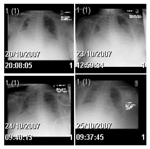
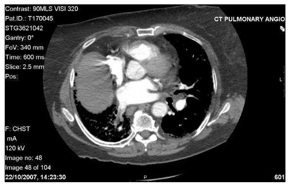
Case presentation: The authors report a case of a 78-year-old previously healthy female presenting with collapse and shortness of breath. Serum biochemistry revealed acute kidney injury, positive D-dimmer's and increased C reactive protein. Chest radiography demonstrated volume loss in the right lung. The patient was started on antibiotics and also therapeutic doses of low molecular weight heparin. The working diagnosis included community acquired pneumonia & pulmonary embolism. A computed tomography pulmonary angiogram was performed to confirm the clinical suspicions of pulmonary embolism. This demonstrated a thrombus in the pulmonary vein, with associated fibrosis and volume loss of the right lower lobe. A subsequent thrombophilia screen revealed a positive lupus anticoagulant antibody and rheumatoid factor and also decreased anti thrombin III and protein C levels. The urine protein/creatinine ratio was found to be 553 mg/mmol.
Conclusion: The diagnosis of this patient was therefore of idiopathic pulmonary fibrosis associated with pulmonary vein thrombosis. Whether or not the pulmonary vein thrombosis was a primary cause of the fibrosis or a consequence of it was unclear. There are few data on the management of pulmonary vein thrombosis, but anticoagulation, antibiotics, and, in cases of large pulmonary vein thrombosis, thrombectomy or pulmonary resection have been used.
Figures
References
-
- Dye TE, Saab SB, Almond CH, Watson L. Sclerosing mediastinitis with occlusion of pulmonary veins: manifestation and management. J Thorac Cardiovasc Surg. 1977;74:137–141. - PubMed
-
- Stevens LH, Hormuth DA, Schmidt PE, Atkins S, Fehrenbacher JW. Left atrial myxoma: pulmonary infarction caused by pulmonary venous occlusion. Ann Thorac Surg. 1987;43:215–217. - PubMed
-
- Julio AM. Pulmonary vein thrombosis presenting as myocardial infarction. Chest. 2006. p. 344S.
LinkOut - more resources
Full Text Sources
Research Materials
