

Imprint cytology of osteosarcoma of the jaw: a case report
- PMID: 20062756
- PMCID: PMC2803850
- DOI: 10.1186/1752-1947-3-9327
Imprint cytology of osteosarcoma of the jaw: a case report
Abstract
Introduction: Osteosarcomas are highly malignant bone-forming neoplasms that account for about 20% of all sarcomas. In light of their aggressive behavior, early diagnosis is crucial for determining adequate treatment. Dental professionals may be the first to detect jaw osteosarcomas in their initial stages. The aim of this case report is to draw attention to the possibility of diagnosing this tumor based on clinical, radiographical and cytological characteristics before confirmation by histology.
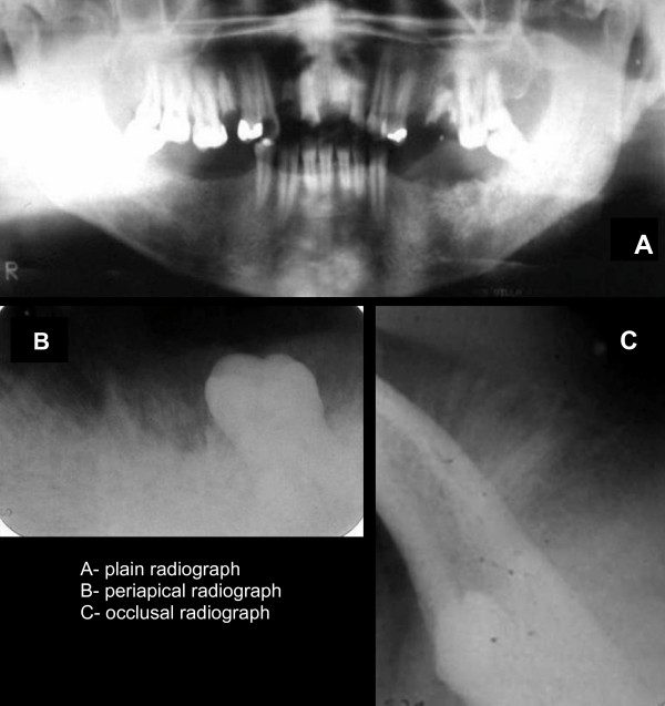
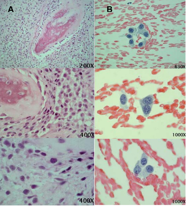
Case presentation: A 24-year-old Afro-Brazilian man presented with swelling and pain on the left side of the mandible in the region of the third molar (tooth 38). Radiography showed a poorly delimited intraosseous lesion with radiolucent and radiopaque areas. The cytological aspects were consistent with the diagnosis of osteosarcoma, which was confirmed by biopsy.
Conclusion: Imprint cytology was found to be a reliable, rapid and easy complementary examination. An early diagnosis of osteosarcoma of the jaw is fundamental to the early determination of an adequate treatment.
Figures


Similar articles
-
Osteosarcoma of Jaw with Varying Histomorphologic Patterns: Case Report.J Orthop Case Rep. 2017 Jan-Feb;7(1):61-64. doi: 10.13107/jocr.2250-0685.690. J Orthop Case Rep. 2017. PMID: 28630843 Free PMC article.
-
Osteosarcoma of the mandible: A case report with an early radiographic manifestation.Imaging Sci Dent. 2014 Mar;44(1):85-8. doi: 10.5624/isd.2014.44.1.85. Epub 2014 Mar 19. Imaging Sci Dent. 2014. PMID: 24701464 Free PMC article.
-
Osteosarcoma metastatic to the mandible: a case report.Oral Surg Oral Med Oral Pathol Oral Radiol Endod. 2001 Apr;91(4):452-4. doi: 10.1067/moe.2001.113107. Oral Surg Oral Med Oral Pathol Oral Radiol Endod. 2001. PMID: 11312462
-
Radiographic features of a patient with both cemento-ossifying fibroma and keratocystic odontogenic tumor in the mandible: a case report and review of literature.Oral Surg Oral Med Oral Pathol Oral Radiol Endod. 2011 Dec;112(6):798-802. doi: 10.1016/j.tripleo.2011.06.025. Epub 2011 Oct 8. Oral Surg Oral Med Oral Pathol Oral Radiol Endod. 2011. PMID: 21986255 Review.
-
Coincidental diagnosis of tuberculous lymphadenitis: a case report.Aust Dent J. 2014 Jun;59(2):258-63. doi: 10.1111/adj.12179. Aust Dent J. 2014. PMID: 24861404 Review.
Cited by
-
Bisphosphonate enhances TRAIL sensitivity to human osteosarcoma cells via death receptor 5 upregulation.Exp Mol Med. 2011 Mar 31;43(3):138-45. doi: 10.3858/emm.2011.43.3.016. Exp Mol Med. 2011. PMID: 21297379 Free PMC article.
-
A Rare Case Report of Osteosarcoma of Maxilla with Double Free-Flap Reconstruction.Indian J Surg Oncol. 2024 Mar;15(Suppl 1):172-178. doi: 10.1007/s13193-023-01846-1. Epub 2023 Nov 21. Indian J Surg Oncol. 2024. PMID: 38545597 Free PMC article.
-
Chondroblastic osteosarcoma of the nasal cavity: an exceptional and misdiagnosed presentation.J Surg Case Rep. 2024 Jun 6;2024(6):rjae409. doi: 10.1093/jscr/rjae409. eCollection 2024 Jun. J Surg Case Rep. 2024. PMID: 38845795 Free PMC article.
References
-
- Schajowicz F. Histological typing of bone tumours. 2. Berlin; New York: Springer-Verlag; 1993. p. 128.
-
- Bennett JH, Thomas G, Evans AW, Speight PM. Osteosarcoma of the jaws: a 30-year retrospective review. Oral Surg Oral Med Oral Pathol Oral Radiol Endod. 2000;90(3):323–332. - PubMed
-
- Chindia ML. Osteosarcoma of the jaw bones. Oral Oncol. 2001;37(7):545–547. - PubMed
-
- Mardinger O, Givol N, Talmi YP, Taicher S. Osteosarcoma of the jaw. The Chaim Sheba Medical Center experience. Oral Surg Oral Med Oral Pathol Oral Radiol Endod. 2001;91(4):445–451. - PubMed
-
- Nakayama E, Sugiura K, Kobayashi I, Oobu K, Ishibashi H, Kanda S. The association between the computed tomography findings, histologic features, and outcome of osteosarcoma of the jaw. J Oral Maxillofac Surg. 2005;63(3):311–318. - PubMed
LinkOut - more resources
Full Text Sources
