

Cecum perforation due to tuberculosis in a renal transplant recipient: a case report
- PMID: 20062770
- PMCID: PMC2803804
- DOI: 10.1186/1752-1947-3-132
Cecum perforation due to tuberculosis in a renal transplant recipient: a case report
Abstract
Introduction: Tuberculosis can present in many varied clinical situations in immunosuppressed patients. It has been reported that the sigmoid colon is the most common site for colonic perforation in renal transplant recipients and diverticulitis is its most common cause. Cecal perforation because of tuberculosis is extremely rare in a renal transplant recipient. We present the case of a renal transplant patient with cecal perforation due to tuberculosis, 10 years after renal transplantation.
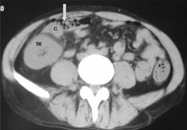
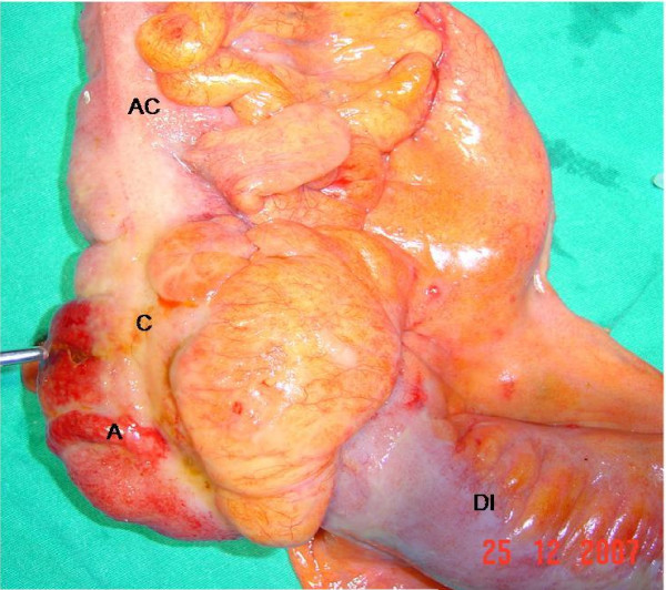
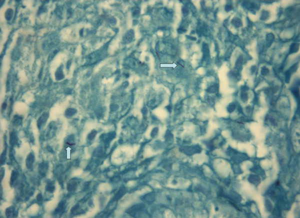
Case presentation: A 39-year-old Caucasian man, who was a renal transplant recipient, was admitted to our emergency surgery unit with an acute abdomen. A cecal perforation was found at exploratory laparotomy, and a right hemicolectomy with an end ileostomy and transverse colonic mucous fistula were performed. Necrotizing granulomatous colitis due to tuberculosis was reported in the histopathologic examination.
Conclusion: Colonic perforations in immunosuppressed patients may have unusual presentations and unusual causes. Tuberculosis infection should be considered in the differential diagnosis during the histopathologic evaluation in immunocompromised patients such as renal transplant recipients.
Figures
Similar articles
-
Sigmoid Colon Tuberculosis Revealed by a Perforation and Peritonitis.Cureus. 2020 Dec 25;12(12):e12272. doi: 10.7759/cureus.12272. Cureus. 2020. PMID: 33520488 Free PMC article.
-
Acute adomen in a transplant patient with tuberculous colitis: a case report.Cases J. 2009 Dec 10;2:9305. doi: 10.1186/1757-1626-2-9305. Cases J. 2009. PMID: 20072674 Free PMC article.
-
Transverse Colonic Perforation in Renal Transplant Recipients During the Early Postoperative Period: A Case Series.Transplant Proc. 2021 Apr;53(3):1070-1074. doi: 10.1016/j.transproceed.2021.01.019. Epub 2021 Feb 8. Transplant Proc. 2021. PMID: 33573821
-
Synchronous diverticular perforation: report of a case.Am Surg. 2005 Jun;71(6):528-31. Am Surg. 2005. PMID: 16044938 Review.
-
Sigmoido-Cecal Fistula: A Rare Case of Complicated Recurrent Diverticulitis and a Review of the Literature.Am J Case Rep. 2018 Nov 22;19:1386-1392. doi: 10.12659/AJCR.911790. Am J Case Rep. 2018. PMID: 30464167 Free PMC article. Review.
Cited by
-
Tuberculous severe acute colitis. A case report.Ann Med Surg (Lond). 2021 Aug 23;69:102756. doi: 10.1016/j.amsu.2021.102756. eCollection 2021 Sep. Ann Med Surg (Lond). 2021. PMID: 34484727 Free PMC article.
-
Primary cecal pathologies presenting as acute abdomen and critical appraisal of their current management strategies in emergency settings with review of literature.Int J Crit Illn Inj Sci. 2018 Apr-Jun;8(2):90-99. doi: 10.4103/IJCIIS.IJCIIS_69_17. Int J Crit Illn Inj Sci. 2018. PMID: 29963412 Free PMC article.
-
Tuberculosis in a renal allograft recipient presenting with intussusception.Indian J Nephrol. 2012 Jan;22(1):52-6. doi: 10.4103/0971-4065.83741. Indian J Nephrol. 2012. PMID: 22279345 Free PMC article.
-
Colon tuberculosis: endoscopic features and prospective endoscopic follow-up after anti-tuberculosis treatment.Clin Transl Gastroenterol. 2012 Oct 11;3(10):e24. doi: 10.1038/ctg.2012.19. Clin Transl Gastroenterol. 2012. PMID: 23238066 Free PMC article.
References
LinkOut - more resources
Full Text Sources
