. 2010 Jan;6(1):e1000717.
doi: 10.1371/journal.ppat.1000717. Epub 2010 Jan 8.

Caenorhabditis elegans N-glycan core beta-galactoside confers sensitivity towards nematotoxic fungal galectin CGL2

Affiliations

Caenorhabditis elegans N-glycan core beta-galactoside confers sensitivity towards nematotoxic fungal galectin CGL2

Alex Butschi et al. PLoS Pathog. 2010 Jan.

Abstract

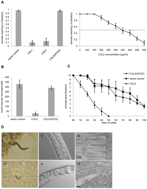
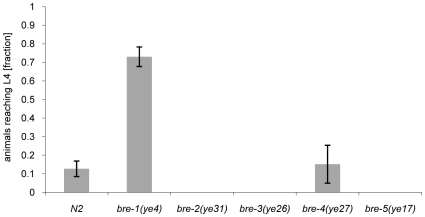
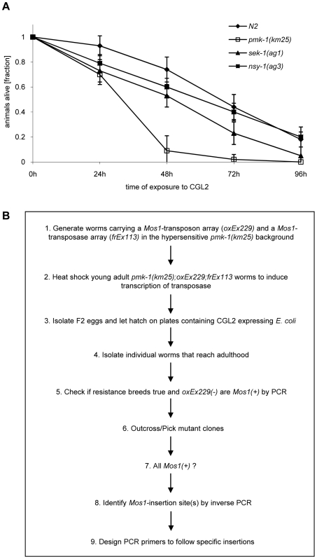
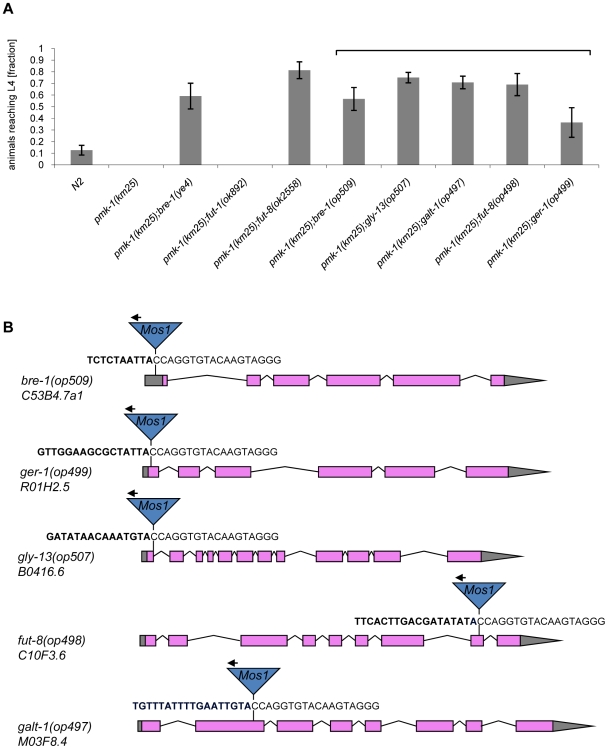
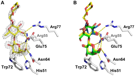
The physiological role of fungal galectins has remained elusive. Here, we show that feeding of a mushroom galectin, Coprinopsis cinerea CGL2, to Caenorhabditis elegans inhibited development and reproduction and ultimately resulted in killing of this nematode. The lack of toxicity of a carbohydrate-binding defective CGL2 variant and the resistance of a C. elegans mutant defective in GDP-fucose biosynthesis suggested that CGL2-mediated nematotoxicity depends on the interaction between the galectin and a fucose-containing glycoconjugate. A screen for CGL2-resistant worm mutants identified this glycoconjugate as a Galbeta1,4Fucalpha1,6 modification of C. elegans N-glycan cores. Analysis of N-glycan structures in wild type and CGL2-resistant nematodes confirmed this finding and allowed the identification of a novel putative glycosyltransferase required for the biosynthesis of this glycoepitope. The X-ray crystal structure of a complex between CGL2 and the Galbeta1,4Fucalpha1,6GlcNAc trisaccharide at 1.5 A resolution revealed the biophysical basis for this interaction. Our results suggest that fungal galectins play a role in the defense of fungi against predators by binding to specific glycoconjugates of these organisms.


Conflict of interest statement

The authors have declared that no competing interests exist.

Figures

Figure 1
Figure 1. Dose and carbohydrate-binding dependent toxicity of C. cinerea galectin CGL2 towards C. elegans.
In all of the experiments, E. coli BL21(DE3) cells expressing either the authentic CGL1 or CGL2 proteins or the carbohydrate-binding defective variant CGL2(W72G), or control transformants were fed to C. elegans wild type N2. (A) CGL2 inhibits C. elegans development. C. elegans L1 larvae were seeded onto lawns of above bacteria (left panel) or fed with increasing concentrations of purified CGL2 together with equal amounts of empty vector-containing BL21(DE3) in liquid culture (right panel) and scored for the fraction developing to the L4 stage within 72 h and 96 h, respectively. Columns represent the average of 6 and 12 replicates, respectively. Error bars indicate standard deviations. The fraction of animals reaching L4 was significantly lower on CGL1- or CGL2-expressing bacteria (p<0.01) than on bacteria expressing CGL2(W72G) or containing empty vector. No significant difference was observed between latter two conditions (p>0.5). In the liquid assay, worm development decreases significantly at CGL2 concentrations higher than 150 µg/ml. (B) CGL2 inhibits C. elegans reproduction. C. elegans hermaphrodites were placed as L4 animals on plates seeded with above bacteria and scored for total progeny counts per hermaphrodite. The broods of eight to ten hermaphrodites were averaged per data point. The standard deviations are indicated. The differences between the L4 fractions on CGL2 and vector control and CGL2(W72G) were statistically significant (p>0.01). None of the progeny on wild type CGL2 developed to adulthood within 96 h post hatch. (C) CGL2 ultimately kills C. elegans. 10 L4 staged wild type C. elegans (N2) were seeded onto lawns of CGL2-, CGL2(W72G)-expressing and empty vector control-containing E. coli BL21(DE3) cells. Each day, the plates were checked for surviving animals which were then transferred to novel bacterial lawns of the same type. The data points represent the average of five replicates. Error bars indicate standard deviations. The survival rate of the worms was significantly impaired on CGL2-expressing bacteria compared to CGL2(W72G) and vector control (p<0.01), whereas there was no significant difference between latter two conditions (p>0.5). (D) CGL2 damages C. elegans intestine. C. elegans L4 larvae were fed with CGL2-expressing (panels i-vi) and control E. coli BL21(DE3) cells (panels i-iii) and examined after 24 h under the stereomicroscope (panels i,iv), by differential interference contrast (DIC) microscopy (panels ii, v) and by transmission electron microscopy (TEM) (panels iii,vi). The size bars in panels iii and vi are 200 nm.
Figure 2
Figure 2. Resistance of bre-1 mutant towards CGL2-mediated toxicity.
L1 larvae of C. elegans bre-1 to bre-5 mutant strains as well as wild-type N2 were seeded onto a lawn of CGL2-expressing E. coli BL21(DE3) cells and scored for the fraction developing to the L4 stage within 72 h. Columns represent the average of 6 replicates. Error bars indicate standard deviations. The fraction of animals reaching L4 was significantly higher for the bre-1 mutant (p<0.01) than for the wild type (N2) or the bre-4 mutant. No significant difference was observed between latter two strains (p>0.5). For the rest of the mutants, not a single larva developed, so results were not compared statistically.
Figure 3
Figure 3. Workflow of the forward genetic screen for CGL2-resistant C. elegans mutants.
(A) CGL2-sensitivity test of pmk-1, sek-1 and nsy-1 mutant worms defective in the p38 MAPK pathway. 10 L4 staged worms of the indicated genotypes were seeded onto a lawn of CGL2-expressing E. coli BL21(DE3) cells. The plates were checked for surviving animals at the indicated time points. The data points represent the average of ten replicates. Error bars indicate standard deviations. The pmk-1 and sek-1 mutants were significantly hypersensitive compared to the N2 wild type strain (p<0.01), whereas the slightly higher sensitivity of the nsy-1 mutant is less significant (p<0.2). (B) Mos1 insertional mutagenesis workflow. Worms that carry the Mos1 transposon array oxEx229 and the Mos1 transposase array frEx113 in the CGL2-hypersensitive pmk-1(km25) background were generated.
Figure 4
Figure 4. Results of the forward genetic screen for CGL2-resistant C. elegans mutants.
(A) Resistance of isolated and constructed C. elegans mutants towards CGL2-mediated toxicity. C. elegans mutants of the indicated genotypes were analysed for development from L1 to L4 as outlined above. The gene appendix (op) and the bracket above the histogram indicate mutants isolated in the Mos1-screen. The other mutants were constructed by crossing. The increase in the fractions of animals reaching L4 of the various mutants compared to N2 worms was statistically significant (p<0.05). In case of the pmk-1 and the pmk-1;fut-1 mutant, not a single larva developed, so results were not compared statistically. (B) Insertion sites of Mos1 elements in CGL2-resistant mutants. Arrows above Mos1 elements indicate the orientation of the Mos1 primer oJL115 used for sequencing iPCR products of mutant lysates. Bold letters indicate C. elegans genomic sequences and are followed by Mos1 sequence. Gene models are taken from WormBase Release WS207. bre-1(op509) mutants have a Mos1 insertion in the 5′-UTR of C53B4.7a1, located 184 bp upstream of the translational start codon. ger-1(op499) mutants have a Mos1 insertion in the first exon of R01H2.5, located 50 bp downstream of the translational start codon. gly-13(op507) mutants have a Mos1 insertion in a conserved splicing donor site flanking the first exon of B0416.6. fut-8(op498) mutants have a Mos1 insertion in the eighth exon of C10F3.6. galt-1(op497) mutants have a Mos1 insertion in the second exon of M03F8.4.
Figure 5
Figure 5. CGL2-sensitivity test and biosynthetic context of available C. elegans fucosyltransferase and GlcNAc-transferase mutants.
(A) CGL2-sensitivity test. C. elegans mutants of the indicated genotypes were analysed for development from L1 to L4 as outlined above. The differences between the L4 fractions of the fut-4(gk111) and dpy-6(e14);gly-13(ok712) mutants to N2 are statistically significant (p<0.01) in contrast to the differences between the L4 fractions of the fut8(ok2558), gly-12(is47) and gly-14(id48) mutants to N2 (p<0.9). The differences of the residual mutants to N2 could not be evaluated due to the lack of variance. (B) Biosynthetic context. The putative pathway for biosynthesis of the CGL2 epitope, based on previous in vivo and in vitro data, indicates the key roles of the enzymes encoded by the gly-13, fut-8 and galt-1(M03F8.4) genes; on the other hand, FUT-1 acts after the processing by hexosaminidases such as HEX-2. Other fucosyltransferases such as FUT-2 through to FUT-6 are not involved in modification of the reducing-terminal GlcNAc residue of N-glycans, whereas the gly-2 and gly-20 encoded enzymes are not prerequisites for α1,6-fucosylation by FUT-8. Further modifications by other enzymes, encoded by unknown genes, are possible and result in structures such as the depicted Hex5dHex2HexNAc2 glycan.
Figure 6
Figure 6. In situ localization of the glycoepitope recognized by CGL2.
C. elegans CGL2-sensitive pmk-1(km25) and CGL2-resistant pmk-1(km25);fut-8(op498) worms were fed with TAMRA-labeled CGL2 and examined by differential interference contrast (DIC) and red fluorescence (RF) microscopy. 32 and 25 animals, respectively, were scored for staining of the intestinal epithelium (see Supplementary Table S1). The numbers of worms with stained intestinal epithelium was significantly different between the two genetic backgrounds (p<0.01).
Figure 7
Figure 7. Comparative analysis of the N-glycome in CGL2-resistant C. elegans double mutants pmk-1;fut-8(op498) (red trace) and pmk-1(km25);M03F8.4(op497) (green trace) and the isogenic CGL2-hypersensitive single mutant strain pmk-1(km25) (black trace).
(A) and (B) HPLC of released and fluorescently labeled N-glycans. Upon enzymatic release (here the PNGase F resistant, but PNGase A sensitive glycome fraction is shown) and fluorescent labeling, N-glycans were separated by normal phase HPLC and analysed by mass spectrometry (A). Fractions at similar retention times (e.g., dashed rectangle) were further separated by reversed phase HPLC and the resulting pure glycans were analysed by mass spectrometry (MS) and for selected fractions by MS/MS (B). (C) Structural characterization of a selected isolated peak. Peak A found in the pmk-1 strain but not in the two double mutant strains was treated with β1,4-galactosidase (β1,4-Gal'ase) and α-fucosidase (α-Fuc'ase). The reaction products were analysed by reversed phase HPLC and MS and MS/MS (see Supplementary Figure S2). The blue HPLC trace represents the glycan standard in glucose units (GU). Monosaccharides are represented as symbols: Man (green circle), Gal (yellow circle), GlcNAc (blue square), Fuc (red triangle), Hex (white circle).
Figure 8
Figure 8. Detailed view of the interaction between CGL2 and Galβ1,4Fucα1,6GlcNAc (A) and comparison with two other CGL2/carbohydrate complexes (B).
(A) Fourier difference map (with Fo - Fc coefficients) around the visible part of ligand contoured at 3 σ. Residues belonging to the binding pocket are displayed as sticks and H-bonds as dashed yellow lines. (B) Superimposition of the lactose (Galβ1,4Glc) (green, PDB ID 1ULC) and of the Thomsen-Friedenreich antigen (Galβ1,3GalNAc) (blue, PDB ID 1ULG) onto the CGL2/Galβ1,4Fucα1,6GlcNAc structure (yellow, PDB ID 2WKK). The binding pocket is almost identical in all three structures.

Similar articles

Cited by

References

    1. Peumans WJ, Van Damme EJ. Lectins as plant defense proteins. Plant Physiol. 1995;109:347–352. - PMC - PubMed
    1. Loris R. Principles of structures of animal and plant lectins. Biochim Biophys Acta. 2002;1572:198–208. - PubMed
    1. Cash HL, Whitham CV, Behrendt CL, Hooper LV. Symbiotic bacteria direct expression of an intestinal bactericidal lectin. Science. 2006;313:1126–1130. - PMC - PubMed
    1. Kohatsu L, Hsu DK, Jegalian AG, Liu FT, Baum LG. Galectin-3 induces death of Candida species expressing specific β1,2-linked mannans. J Immunol. 2006;177:4718–4726. - PubMed
    1. Griffitts JS, Haslam SM, Yang T, Garczynski SF, Mulloy B, et al. Glycolipids as receptors for Bacillus thuringiensis crystal toxin. Science. 2005;307:922–925. - PubMed

Publication types

MeSH terms