. 2010 Nov;39(11):1103-8.
doi: 10.1007/s00256-009-0857-9. Epub 2010 Jan 9.

Measuring hindfoot alignment radiographically: the long axial view is more reliable than the hindfoot alignment view

Affiliations

Measuring hindfoot alignment radiographically: the long axial view is more reliable than the hindfoot alignment view

Mikel L Reilingh et al. Skeletal Radiol. 2010 Nov.

Abstract

Background: Hindfoot malalignment is a recognized cause of foot and ankle disability. For preoperative planning and clinical follow-up, reliable radiographic assessment of hindfoot alignment is important. The long axial radiographic view and the hindfoot alignment view are commonly used for this purpose. However, their comparative reliabilities are unknown. As hindfoot varus or valgus malalignment is most pronounced during mid-stance of gait, a unilateral weight-bearing stance, in comparison with a bilateral stance, could increase measurement reliability. The purpose of this study was to compare the intra- and interobserver reliability of hindfoot alignment measurements of both radiographic views in bilateral and unilateral stance.

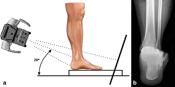
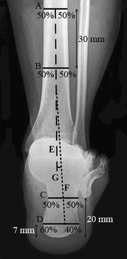
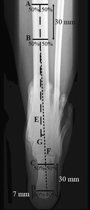
Materials and methods: A hindfoot alignment view and a long axial view were acquired from 18 healthy volunteers in bilateral and unilateral weight-bearing stances. Hindfoot alignment was defined as the angular deviation between the tibial anatomical axis and the calcaneus longitudinal axis from the radiographs. Repeat measurements of hindfoot alignment were performed by nine orthopaedic examiners.

Results: Measurements from the hindfoot alignment view gave intra- and interclass correlation coefficients (CCs) of 0.72 and 0.58, respectively, for bilateral stance and 0.91 and 0.49, respectively, for unilateral stance. The long axial view showed, respectively, intra- and interclass CCs of 0.93 and 0.79 for bilateral stance and 0.91 and 0.58 for unilateral stance.

Conclusion: The long axial view is more reliable than the hindfoot alignment view or the angular measurement of hindfoot alignment. Although intra-observer reliability is good/excellent for both methods, only the long axial view leads to good interobserver reliability. A unilateral weight-bearing stance does not lead to greater reliability of measurement.


Figures

Fig. 1
Fig. 1
a The hindfoot alignment view. The inclination angle of the beam is 20° to the floor. The film cassette is perpendicular to the central beam of the radiation source. b A random example of a radiograph showing the hindfoot alignment view
Fig. 2
Fig. 2
a The long axial view. The film cassette is lying on the floor and the subject is standing on the film cassette. The inclination angle of the beam is 45° to the floor. b A random example of a radiograph showing the long axial view
Fig. 3
Fig. 3
Measurement method for the hindfoot alignment view. We defined the mid-diaphyseal axis of the tibia by bisecting the tibia into two mid-diaphyseal points (lines A and B) 30 mm apart and extending the line distally (line E). The mid-diaphyseal axis of the calcaneus is defined by a line through two points in the calcaneus. At a distance of 7 mm from the most distal part of the calcaneus, a horizontal line is drawn (line D). Line D is divided into a 40%:60% ratio, where the length of the 40% line is measured from the lateral side. A second line (line C) is drawn horizontally, 20 mm from the most distal part of the calcaneus. The calcaneus axis (line F) is drawn by connecting the 40% mark at line D and the bisected line C. The hindfoot angle (G) is the angle between lines E and F
Fig. 4
Fig. 4
Measurement method for the long axial view. We defined the mid-diaphyseal axis of the tibia by bisecting the tibia into two mid-diaphyseal points (lines A and B) 30 mm apart and extending the line distally (line E). The mid-diaphyseal axis of the calcaneus is defined by a line through two points in the calcaneus. At a distance of 7 mm from the most distal part of the calcaneus, a horizontal line is drawn (line D). Line D is divided into a 40%:60% ratio, where the length of the 40% line is measured from the lateral side. A second line (line C) is drawn horizontally, 30 mm from the most distal part of the calcaneus. The calcaneus axis (line F) is drawn by connecting the 40% mark at line D and the bisected line C. The hindfoot angle (G) is the angle between lines E and F

References

    1. Hayashi K, Tanaka Y, Kumai T, Sugimoto K, Takakura Y. Correlation of compensatory alignment of the subtalar joint to the progression of primary osteoarthritis of the ankle. Foot Ankle Int. 2008;29:400–406. doi: 10.3113/FAI.2008.0400. - DOI - PubMed
    1. Takakura Y, Takaoka T, Tanaka Y, Yajima H, Tamai S. Results of opening-wedge osteotomy for the treatment of a post-traumatic varus deformity of the ankle. J Bone Jt Surg Am. 1998;80:213–218. - PubMed
    1. Hintermann B, Knupp M, Barg A. Osteotomies of the distal tibia and hindfoot for ankle realignment. Orthopade. 2008;37:212–213. doi: 10.1007/s00132-008-1214-8. - DOI - PubMed
    1. Stamatis ED, Myerson MS. Supramalleolar osteotomy: indications and technique. Foot Ankle Clin. 2003;8:317–333. doi: 10.1016/S1083-7515(03)00018-4. - DOI - PubMed
    1. Tanaka Y, Takakura Y, Hayashi K, Taniguchi A, Kumai T, Sugimoto K. Low tibial osteotomy for varus-type osteoarthritis of the ankle. J Bone Joint Surg Br. 2006;88:909–913. doi: 10.2106/JBJS.E.00472. - DOI - PubMed