. 2010 Feb;34(2):311-6.
doi: 10.1007/s00264-009-0933-8. Epub 2010 Jan 9.

The sulcus deepening trochleoplasty-the Lyon's procedure

Affiliations

The sulcus deepening trochleoplasty-the Lyon's procedure

David Dejour et al. Int Orthop. 2010 Feb.

Abstract

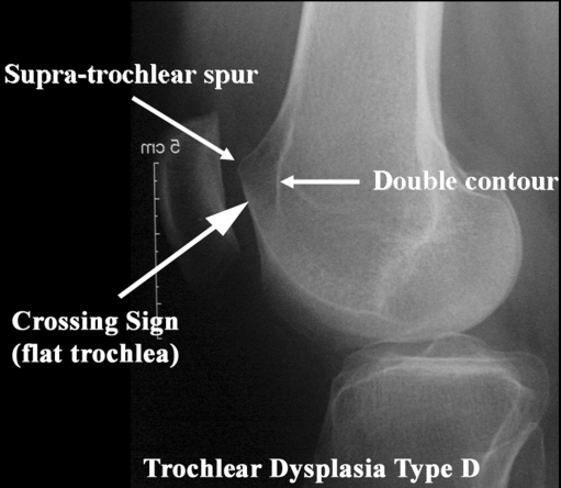
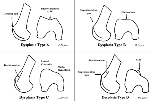
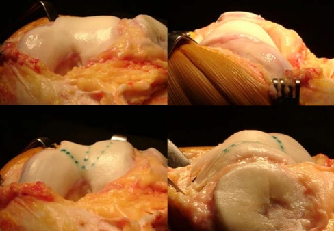
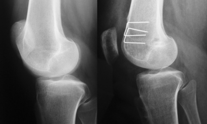
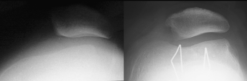
Sulcus deepening trochleoplasty is a technically demanding procedure with precise indications: high grade trochlear dysplasia with patellar instability and/or abnormal tracking. The main goal is to decrease the prominence of the trochlea and to create a new groove with normal depth, thus optimising patellar tracking. Associated abnormalities should be specifically treated. Recurrence of instability is very rare after this procedure and is more likely to result from missed associated abnormalities. Although results seem very good in terms of instability, further evidence is still needed since the groups of patients in the published series are heterogeneous. Trochleoplasty is not indicated for patellofemoral arthritis or pain. As any surgical procedure, sulcus deepening trochleoplasty is liable to complications.


Figures

Fig. 1
Fig. 1
To analyse trochlear dysplasia a true profile is needed with a perfect superimposition of the posterior condyles. The three trochlear dysplasia signs are: the crossing sign, the supra-trochlear spur and the double-contour (which appears below the crossing sign)
Fig. 2
Fig. 2
Trochlear dysplasia classification according to D. Dejour. In type A, there is the crossing sign is present in lateral true view. The trochlea is shallower than normal, but still symmetrical and concave. Type B features include the crossing sign and trochlear spur. The trochlea is flat or convex in axial images. In type C there is a crossing sign, and in addition the double-contour sign representing sclerosis of the subchondral bone of the medial hypoplastic facet. On axial CT scan views, the lateral facet is convex. Type D combines all the mentioned signs: crossing sign, supratrochlear spur and double-contour sign going below the crossing sign. In axial CT scan views, there is a cliff pattern
Fig. 3
Fig. 3
Drawing of the new trochlear limits; the medial facet is nearly nonexistent
Fig. 4
Fig. 4
Sulcus deepening trochleoplasty schematic model. Cancellous bone from the trochlear undersurface is removed to allow modelling (gray zone). The bone–cartilage flap is then modelled, and fixation is achieved with staples
Fig. 5
Fig. 5
Trochlear shape before (above) and after (below) sulcus deepening trochleoplasty. The trochlear sulcus is restored and more “anatomical” shape is achieved
Fig. 6
Fig. 6
Sulcus deepening trochleoplasty: pre and postoperative lateral views. The supratrochlear spur resection can be observed and the patellar tilt is corrected
Fig. 7
Fig. 7
Sulcus deepening trochleoplasty: pre and postoperative axial views. The sulcus is almost absent in the preoperative view, and restored in the postoperative. Patellar tilt and subluxation are corrected

Similar articles

Cited by

References

    1. Amis AA. Current concepts on anatomy and biomechanics of patellar stability. Sports Med Arthrosc. 2007;15:48–56. doi: 10.1097/JSA.0b013e318053eb74. - DOI - PubMed
    1. Shih YF, Bull AM, Amis AA. The cartilaginous and osseous geometry of the femoral trochlear groove. Knee Surg Sports Traumatol Arthrosc. 2004;12:300–306. doi: 10.1007/s00167-003-0414-3. - DOI - PubMed
    1. Dejour H, Walch G, Neyret P, Adeleine P. Dysplasia of the femoral trochlea. Rev Chir Orthop Reparatrice Appar Mot. 1990;76:45–54. - PubMed
    1. Dejour H, Walch G, Nove-Josserand L, Guier C. Factors of patellar instability: an anatomic radiographic study. Knee Surg Sports Traumatol Arthrosc. 1994;2:19–26. doi: 10.1007/BF01552649. - DOI - PubMed
    1. Tavernier T, Dejour D. Knee imaging: what is the best modality. J Radiol. 2001;82(387–405):407–388. - PubMed