

Chronic social isolation compromises the activity of both glutathione peroxidase and catalase in hippocampus of male wistar rats
- PMID: 20063054
- PMCID: PMC11498870
- DOI: 10.1007/s10571-009-9493-0
Chronic social isolation compromises the activity of both glutathione peroxidase and catalase in hippocampus of male wistar rats
Abstract
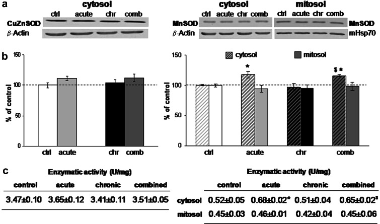
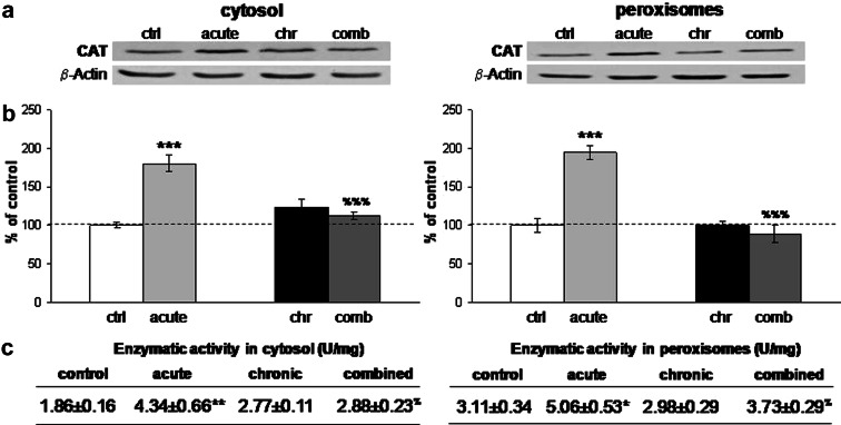
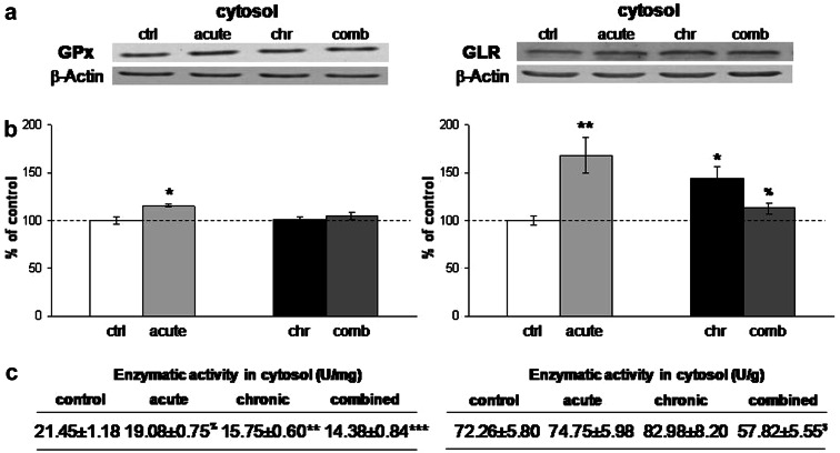
Chronic neuroendocrine stress usually leads to the elevation of the stress hormones and increased metabolic rate, which is frequently accompanied by oxidative damage to the CNS. In the present study we hypothesized that chronic psychosocial isolation (CPSI) of male Wistar rats, characterized by decreased serum corticosterone (CORT), unaltered catecholamines (CTs), and low blood glucose (GLU), may also promote oxidative imbalance in the CNS, by targeting antioxidant defense system. To test it, we have examined the relation between these input signals and protein expression/activity of antioxidant enzymes (AOEs): superoxide dismutases (SODs), catalase (CAT), glutathione peroxidase (GPx), and glutathione reductase (GLR) in the hippocampus (HIPPO) of CPSI animals. We found that CPSI did not affect SODs or CAT, but decreased activity of GPx and compromised GLR, an enzyme highly dependent on blood GLU for its substrate precursor. Further, we have tested whether the CPSI experience altered AOEs response to a novelty stress, and found that it attenuated peroxide-metabolizing enzymes, CAT and GPx, and decreased GLR activity, even though blood GLU was restored. The altered ratios of hippocampal AOEs in CPSI animals, which were worsened under the combined stress conditions, may lead to the accumulation of peroxide products and oxidative imbalance. The mechanism by which CPSI generate oxidative imbalance in the HIPPO is most likely based on poor systemic energy conditions set by this stress. Such conditions may cause functional decline of CNS structures, such as HIPPO, and are likely to promote state linked to onset of many mood disorders.
Figures
Similar articles
-
Chronic stress differentially affects antioxidant enzymes and modifies the acute stress response in liver of Wistar rats.Physiol Res. 2010;59(5):729-736. doi: 10.33549/physiolres.931862. Epub 2010 Apr 20. Physiol Res. 2010. PMID: 20406049
-
Modulation of Hippocampal Antioxidant Defense System in Chronically Stressed Rats by Lithium.Oxid Med Cell Longev. 2019 Feb 17;2019:8745376. doi: 10.1155/2019/8745376. eCollection 2019. Oxid Med Cell Longev. 2019. PMID: 30911352 Free PMC article.
-
[Age-dependent changes of superoxide dismutase, glutathione peroxidase, and catalase activity in the brain of rats during immobilized stress].Ukr Biokhim Zh (1999). 2004 May-Jun;76(3):74-8. Ukr Biokhim Zh (1999). 2004. PMID: 19621742 Russian.
-
Lead-induced dysregulation of superoxide dismutases, catalase, glutathione peroxidase, and guanylate cyclase.Environ Res. 2005 May;98(1):33-9. doi: 10.1016/j.envres.2004.05.016. Environ Res. 2005. PMID: 15721881
-
Impact of an acute exposure to ethanol on the oxidative stress status in the hippocampus of prenatal restraint stress adolescent male rats.Brain Res. 2008 Jan 29;1191:55-62. doi: 10.1016/j.brainres.2007.11.031. Epub 2007 Nov 28. Brain Res. 2008. PMID: 18096141
Cited by
-
Severe life stress and oxidative stress in the brain: from animal models to human pathology.Antioxid Redox Signal. 2013 Apr 20;18(12):1475-90. doi: 10.1089/ars.2012.4720. Epub 2012 Aug 6. Antioxid Redox Signal. 2013. PMID: 22746161 Free PMC article. Review.
-
Effects of Fructose and Stress on Rat Renal Copper Metabolism and Antioxidant Enzymes Function.Int J Mol Sci. 2022 Aug 12;23(16):9023. doi: 10.3390/ijms23169023. Int J Mol Sci. 2022. PMID: 36012287 Free PMC article.
-
Effects of Liquid Fructose Supplementation and Chronic Unpredictable Stress on Uterine Contractile Activity in Nonpregnant Rats.Int J Mol Sci. 2024 Jun 20;25(12):6770. doi: 10.3390/ijms25126770. Int J Mol Sci. 2024. PMID: 38928475 Free PMC article.
-
Chronic Variable Stress Is Responsible for Lipid and DNA Oxidative Disorders and Activation of Oxidative Stress Response Genes in the Brain of Rats.Oxid Med Cell Longev. 2017;2017:7313090. doi: 10.1155/2017/7313090. Epub 2017 Sep 11. Oxid Med Cell Longev. 2017. PMID: 29085557 Free PMC article.
-
Oxidative Dysregulation in Early Life Stress and Posttraumatic Stress Disorder: A Comprehensive Review.Brain Sci. 2021 May 29;11(6):723. doi: 10.3390/brainsci11060723. Brain Sci. 2021. PMID: 34072322 Free PMC article. Review.
References
-
- Adzic M, Djordjevic J, Djordjevic A, Niciforovic A, Demonacos C, Radojcic MB, Krstic-Demonacos M (2009b) Acute or chronic stress induce cell compartment-specific phosphorylation of glucocorticoid receptor and alter its transcriptional activity in Wistar rat brain. J Endocrinol 202(1):87–97 - PMC - PubMed
-
- Barnham KJ, Masters CL, Bush AI (2004) Neurodegenerative diseases and oxidative stress. Nat Rev Drug Discov 3(3):205–214 - PubMed
-
- Bruce AJ, Boling W, Kindy MS, Peschon J, Kraemer PJ, Carpenter MK, Holtsberg FW, Mattson MP (1996) Altered neuronal and microglial responses to excitotoxic and ischemic brain injury in mice lacking TNF receptors. Nat Med 2(7):788–794 - PubMed
-
- Claiborne A (1985) Handbook of methods for oxygene radical research. CRC Press, Boca Raton, FL, pp 283–284
Publication types
MeSH terms
Substances
LinkOut - more resources
Full Text Sources
Research Materials
Miscellaneous
