

Assembly of bacteriophage P2 capsids from capsid protein fused to internal scaffolding protein
- PMID: 20063181
- PMCID: PMC2848670
- DOI: 10.1007/s11262-009-0442-2
Assembly of bacteriophage P2 capsids from capsid protein fused to internal scaffolding protein
Abstract
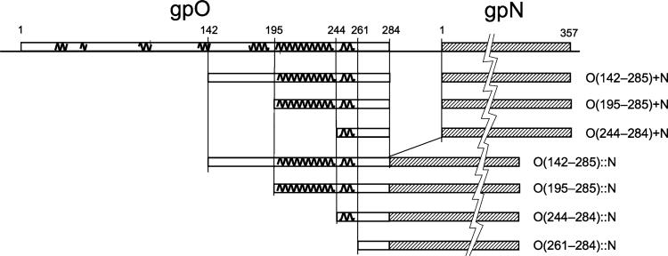
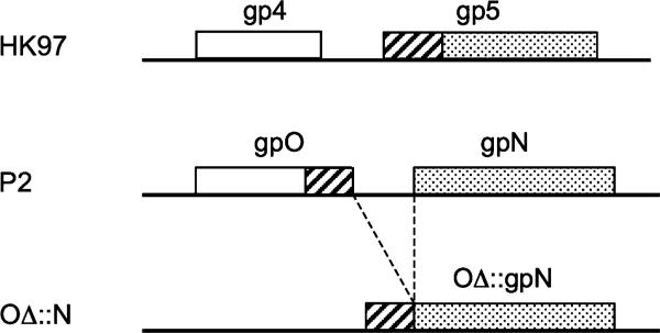
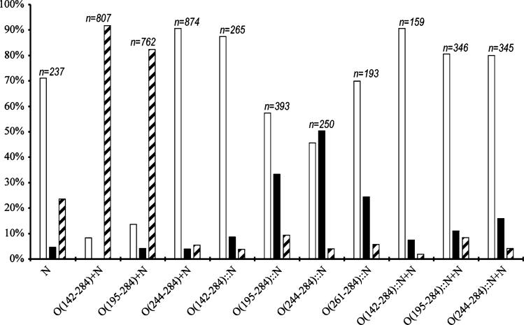
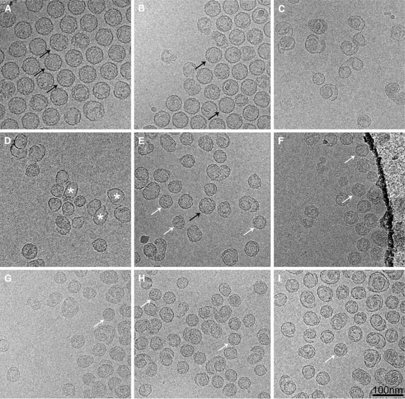
Most tailed bacteriophages with double-stranded DNA genomes code for a scaffolding protein, which is required for capsid assembly, but is removed during capsid maturation and DNA packaging. The gpO scaffolding protein of bacteriophage P2 also doubles as a maturation protease, while the scaffolding activity is confined to a 90 residue C-terminal "scaffolding" domain. Bacteriophage HK97 lacks a separate scaffolding protein; instead, an N-terminal "delta" domain in the capsid protein appears to serve an analogous role. We asked whether the C-terminal scaffolding domain of gpO could work as a delta domain when fused to the gpN capsid protein. Varying lengths of C-terminal sequences from gpO were fused to the N-terminus of gpN and expressed in E. coli. The presence of just the 41 C-terminal residues of gpO increased the fidelity of assembly and promoted the formation of closed shells, but the shells formed were predominantly small, 40 nm shells, compared to the normal, 55 nm P2 procapsid shells. Larger scaffolding domains fused to gpN caused the formation of shells of varying size and shape. The results suggest that while fusing the scaffolding protein to the capsid protein assists in shell closure, it also restricts the conformational variability of the capsid protein.
Keywords: Assembly; Cryo-electron microscopy; Procapsid; Size determination; Virus.
Figures
Similar articles
-
Incorporation of scaffolding protein gpO in bacteriophages P2 and P4.Virology. 2008 Jan 20;370(2):352-61. doi: 10.1016/j.virol.2007.08.039. Epub 2007 Nov 1. Virology. 2008. PMID: 17931675 Free PMC article.
-
Assembly of bacteriophage P2 and P4 procapsids with internal scaffolding protein.Virology. 2006 Apr 25;348(1):133-40. doi: 10.1016/j.virol.2005.12.021. Epub 2006 Feb 7. Virology. 2006. PMID: 16457867
-
Functional domains of the bacteriophage P2 scaffolding protein: identification of residues involved in assembly and protease activity.Virology. 2009 Feb 5;384(1):144-50. doi: 10.1016/j.virol.2008.11.016. Epub 2008 Dec 6. Virology. 2009. PMID: 19064277 Free PMC article.
-
Bacteriophage HK97 capsid assembly and maturation.Adv Exp Med Biol. 2012;726:351-63. doi: 10.1007/978-1-4614-0980-9_15. Adv Exp Med Biol. 2012. PMID: 22297521 Review.
-
Nature's favorite building block: Deciphering folding and capsid assembly of proteins with the HK97-fold.Virology. 2015 May;479-480:487-97. doi: 10.1016/j.virol.2015.02.055. Epub 2015 Apr 8. Virology. 2015. PMID: 25864106 Free PMC article. Review.
Cited by
-
Convergent evolution of pathogenicity islands in helper cos phage interference.Philos Trans R Soc Lond B Biol Sci. 2016 Nov 5;371(1707):20150505. doi: 10.1098/rstb.2015.0505. Philos Trans R Soc Lond B Biol Sci. 2016. PMID: 27672154 Free PMC article.
-
Competing scaffolding proteins determine capsid size during mobilization of Staphylococcus aureus pathogenicity islands.Elife. 2017 Oct 6;6:e30822. doi: 10.7554/eLife.30822. Elife. 2017. PMID: 28984245 Free PMC article.
References
-
- Fane BA, Prevelige PE. Mechanism of scaffolding-assisted viral assembly. Adv. Protein Chem. 2003;64:259–299. - PubMed
-
- Marvik OJ, Sharma P, Dokland T, Lindqvist BH. Bacteriophage P2 and P4 assembly: alternative scaffolding proteins regulate capsid size. Virology. 1994;200:702–714. - PubMed
-
- Wang S, Chang JR, Dokland T. Assembly of bacteriophage P2 and P4 procapsids with internal scaffolding protein. Virology. 2006;348:133–140. - PubMed
Publication types
MeSH terms
Substances
Grants and funding
LinkOut - more resources
Full Text Sources
