

Functional response in ventral temporal cortex differentiates mild cognitive impairment from normal aging
- PMID: 20063353
- PMCID: PMC3004147
- DOI: 10.1002/hbm.20932
Functional response in ventral temporal cortex differentiates mild cognitive impairment from normal aging
Abstract
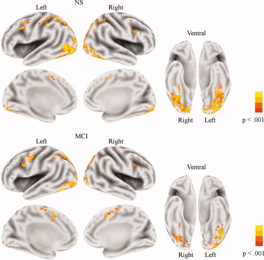
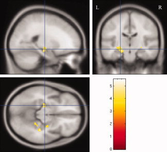
This study sought to identify altered brain activation patterns in amnestic mild cognitive impairment (MCI) that could precede frank task impairment and neocortical atrophy. A high-accuracy lexical decision (LD) task was therefore employed. Both MCI and normal seniors (NS) groups completed the LD task while functional magnetic resonance imaging (fMRI) was performed. Accuracy on the LD task was high (> or =89% correct for both groups), and both groups activated a network of occipitotemporal regions and inferior frontal cortex. However, compared with the NS group, the MCI group showed reduced fMRI activation in these regions and increased activation in bilateral portions of anterior cingluate cortex. The results from a voxel-based morphometry analysis indicated that altered activations in the MCI group were not within regions of atrophy. Receiver operating characteristic curves demonstrated that reduced fMRI response in the left and right midfusiform gyri accurately discriminated MCI from NS. When activation magnitude in both fusiform gyri were included in a single logistic regression model, group classification accuracy was very high (area under the curve = 0.93). These results showed that a disrupted functional response in the ventral temporal lobe accurately distinguishes individuals with MCI from NS, a finding which may have implications for identifying seniors at risk for cognitive decline.
2010 Wiley-Liss, Inc.
Figures


Similar articles
-
Differential cortical atrophy in subgroups of mild cognitive impairment.Arch Neurol. 2005 Sep;62(9):1393-7. doi: 10.1001/archneur.62.9.1393. Arch Neurol. 2005. PMID: 16157746
-
Functional connectivity of the fusiform gyrus during a face-matching task in subjects with mild cognitive impairment.Brain. 2006 May;129(Pt 5):1113-24. doi: 10.1093/brain/awl051. Epub 2006 Mar 6. Brain. 2006. PMID: 16520329
-
Event-related functional magnetic resonance imaging investigation of executive control in very old individuals with mild cognitive impairment.Biol Psychiatry. 2005 Apr 1;57(7):761-7. doi: 10.1016/j.biopsych.2004.12.031. Biol Psychiatry. 2005. PMID: 15820233 Free PMC article.
-
Structural magnetic resonance imaging for the early diagnosis of dementia due to Alzheimer's disease in people with mild cognitive impairment.Cochrane Database Syst Rev. 2020 Mar 2;3(3):CD009628. doi: 10.1002/14651858.CD009628.pub2. Cochrane Database Syst Rev. 2020. PMID: 32119112 Free PMC article.
-
Structural and functional MRI in mild cognitive impairment.Curr Alzheimer Res. 2009 Apr;6(2):179-85. doi: 10.2174/156720509787602898. Curr Alzheimer Res. 2009. PMID: 19355853 Review.
Cited by
-
Estimation of Brain Functional Connectivity in Patients with Mild Cognitive Impairment.Brain Sci. 2019 Nov 30;9(12):350. doi: 10.3390/brainsci9120350. Brain Sci. 2019. PMID: 31801260 Free PMC article.
-
Lifelong bilingualism contributes to cognitive reserve against white matter integrity declines in aging.Neuropsychologia. 2013 Nov;51(13):2841-6. doi: 10.1016/j.neuropsychologia.2013.09.037. Epub 2013 Oct 5. Neuropsychologia. 2013. PMID: 24103400 Free PMC article.
-
A cognitive electrophysiological signature differentiates amnestic mild cognitive impairment from normal aging.Alzheimers Res Ther. 2017 Jan 19;9(1):3. doi: 10.1186/s13195-016-0229-3. Alzheimers Res Ther. 2017. PMID: 28100252 Free PMC article.
-
Staging neurodegenerative disorders: structural, regional, biomarker, and functional progressions.Neurotox Res. 2011 Feb;19(2):211-34. doi: 10.1007/s12640-010-9190-2. Neurotox Res. 2011. PMID: 20393891 Review.
-
How does decisional capacity evolve with normal cognitive aging: systematic review of the literature.Eur Geriatr Med. 2020 Feb;11(1):117-129. doi: 10.1007/s41999-019-00251-8. Epub 2019 Oct 12. Eur Geriatr Med. 2020. PMID: 32297227
References
-
- Ashburner J, Friston KJ ( 2005): Unified segmentation. Neuroimage 26: 839–851. - PubMed
-
- Brun A, Englund E ( 1981): Regional pattern of degeneration in Alzheimer's disease: Neuronal loss and histopathologic grading. Histopathology 5: 549–564. - PubMed
-
- Buchel C, Price C, Friston K ( 1996): A multimodal language region in the ventral visual pathway. Nature 394: 274–277. - PubMed
-
- Busatto GF, Garrido GE, Almeida OP, Castro CC, Camargo CH, Cid CG, Buchpiguel CA, Furuie S, Bottino CM ( 2003): A voxel‐based morphometry study of temporal lobe gray matter reductions in Alzheimer's disease. Neurobiol Aging 24: 22–31. - PubMed
-
- Bush ALH, Allen PA, Kaut KP, Ogrocki PK ( 2007): Influence of mild cognitive impairment on visual word recognition. Aging Neuropsychol Cogn 14: 329–352. - PubMed
Publication types
MeSH terms
Substances
Grants and funding
LinkOut - more resources
Full Text Sources
Medical
Research Materials
Miscellaneous
