Measuring sparseness in the brain: comment on Bowers (2009)
- PMID: 20063978
- PMCID: PMC3154835
- DOI: 10.1037/a0016917
Measuring sparseness in the brain: comment on Bowers (2009)
Abstract
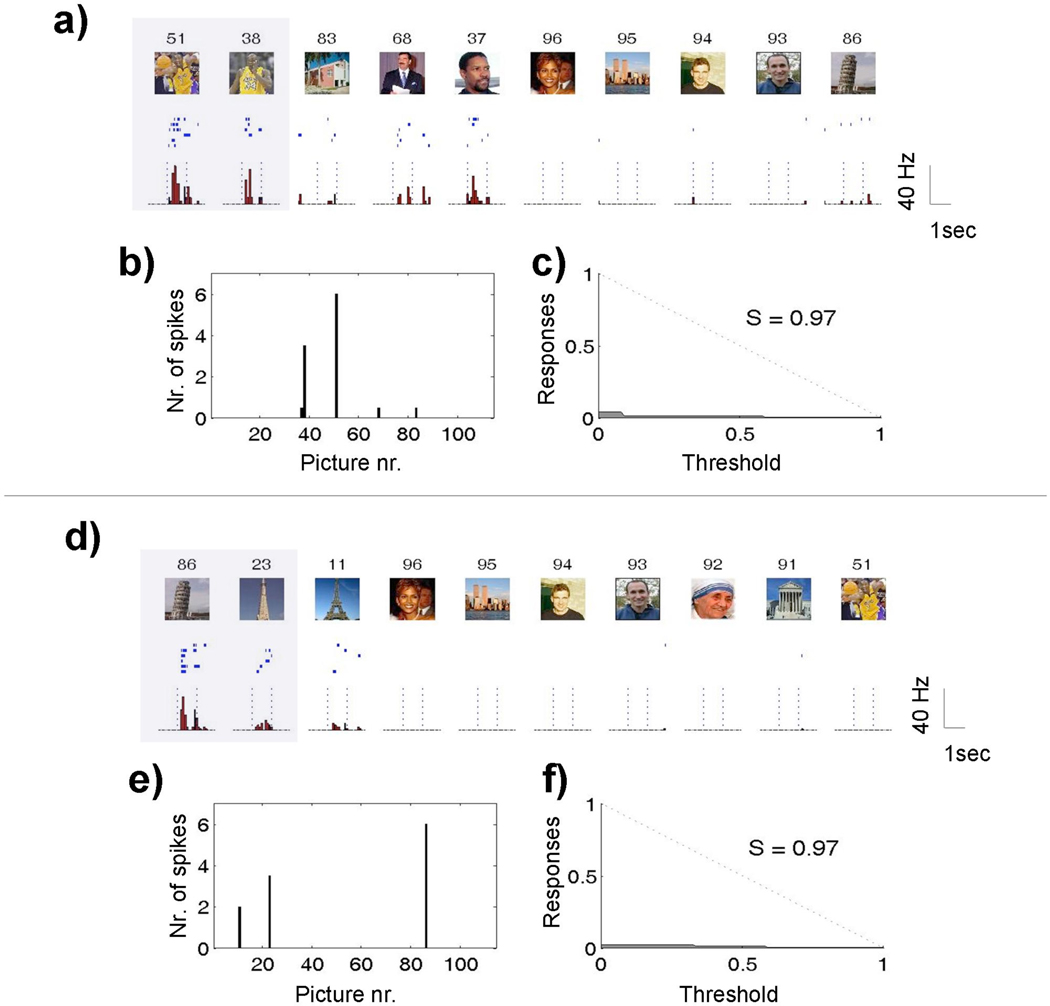
Bowers challenged the common view in favor of distributed representations in psychological modeling and the main arguments given against localist and grandmother cell coding schemes. He revisited the results of several single-cell studies, arguing that they do not support distributed representations. We praise the contribution of Bowers (2009) for joining evidence from psychological modeling and neurophysiological recordings, but we disagree with several of his claims. In this comment, we argue that distinctions between distributed, localist, and grandmother cell coding can be troublesome with real data. Moreover, these distinctions seem to be lying within the same continuum, and we argue that it may be sensible to characterize coding schemes with a sparseness measure. We further argue that there may not be a unique coding scheme implemented in all brain areas and for all possible functions. In particular, current evidence suggests that the brain may use distributed codes in primary sensory areas and sparser and invariant representations in higher areas.
Figures
Comment on
-
On the biological plausibility of grandmother cells: implications for neural network theories in psychology and neuroscience.Psychol Rev. 2009 Jan;116(1):220-51. doi: 10.1037/a0014462. Psychol Rev. 2009. PMID: 19159155
Similar articles
-
Locating object knowledge in the brain: comment on Bowers's (2009) attempt to revive the grandmother cell hypothesis.Psychol Rev. 2010 Jan;117(1):284-8. doi: 10.1037/a0017101. Psychol Rev. 2010. PMID: 20063976 Review.
-
On the biological plausibility of grandmother cells: implications for neural network theories in psychology and neuroscience.Psychol Rev. 2009 Jan;116(1):220-51. doi: 10.1037/a0014462. Psychol Rev. 2009. PMID: 19159155
-
Neuronal codes for visual perception and memory.Neuropsychologia. 2016 Mar;83:227-241. doi: 10.1016/j.neuropsychologia.2015.12.016. Epub 2015 Dec 18. Neuropsychologia. 2016. PMID: 26707718 Review.
-
Discrete capacity limits in visual working memory.Curr Opin Neurobiol. 2010 Apr;20(2):177-82. doi: 10.1016/j.conb.2010.03.005. Epub 2010 Mar 31. Curr Opin Neurobiol. 2010. PMID: 20362427 Free PMC article. Review.
-
The neuronal basis of visual memory and imagery in the primate: a neurophysiological approach.Adv Biophys. 1998;35:103-19. Adv Biophys. 1998. PMID: 9949767 Review.
Cited by
-
Neuromorphic artificial intelligence systems.Front Neurosci. 2022 Sep 14;16:959626. doi: 10.3389/fnins.2022.959626. eCollection 2022. Front Neurosci. 2022. PMID: 36188479 Free PMC article. Review.
-
A multilevel account of hippocampal function in spatial and concept learning: Bridging models of behavior and neural assemblies.Sci Adv. 2023 Jul 21;9(29):eade6903. doi: 10.1126/sciadv.ade6903. Epub 2023 Jul 21. Sci Adv. 2023. PMID: 37478189 Free PMC article.
-
Neuronal mechanisms for sequential activation of memory items: Dynamics and reliability.PLoS One. 2020 Apr 16;15(4):e0231165. doi: 10.1371/journal.pone.0231165. eCollection 2020. PLoS One. 2020. PMID: 32298290 Free PMC article.
-
A theory of the brain: localist representation is used widely in the brain.Front Psychol. 2012 Dec 4;3:551. doi: 10.3389/fpsyg.2012.00551. eCollection 2012. Front Psychol. 2012. PMID: 23426117 Free PMC article. No abstract available.
-
Dense and Persistent Odor Representations in the Olfactory Bulb of Awake Mice.J Neurosci. 2024 Sep 25;44(39):e0116242024. doi: 10.1523/JNEUROSCI.0116-24.2024. J Neurosci. 2024. PMID: 39187379 Free PMC article.
References
-
- Abbott LF. Decoding neuronal firing and modelling neural networks. Q Rev Biophys. 1994;27(3):291–331. - PubMed
-
- Abbott LF, Rolls ET, Tovee MJ. Representational capacity of face coding in monkeys. Cereb Cortex. 1996;6:498–505. - PubMed
-
- Barnes CA, McNaughton BL, Mizumori SJY, Leonard BW, Lin L-H. Comparison of spatial and temporal characteristics of neuronal activity in sequential stages of hippocampal processing. Progress in Brain Research. 1990;83:287–300. - PubMed
-
- Bowers JS. On the biological plausibility of grandmother cells: Implications for neural network theories in psychology and neuroscience. Psychological Review. 2009;116(1):220–251. - PubMed
-
- Connor CE, Brincat SL, Pasupathy A. Transformation of shape information in the ventral pathway. Current Opinion in Neurobiology. 2007;17:140–147. - PubMed