

Amifostine reduces the seminiferous epithelium damage in doxorubicin-treated prepubertal rats without improving the fertility status
- PMID: 20064221
- PMCID: PMC2832784
- DOI: 10.1186/1477-7827-8-3
Amifostine reduces the seminiferous epithelium damage in doxorubicin-treated prepubertal rats without improving the fertility status
Abstract
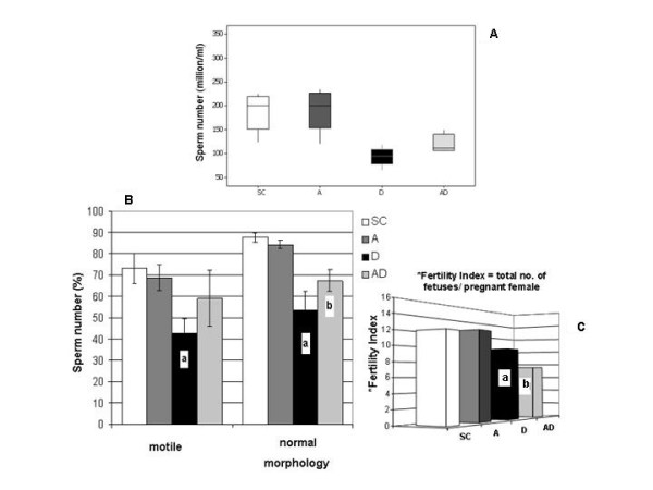
Background: Amifostine is an efficient cytoprotector against toxicity caused by some chemotherapeutic drugs. Doxorubicin, a potent anticancer anthracycline, is known to produce spermatogenic damage even in low doses. Although some studies have suggested that amifostine does not confer protection to doxorubicin-induced testicular damage, schedules and age of treatment have different approach depending on the protocol. Thus, we proposed to investigate the potential cytoprotective action of amifostine against the damage provoked by doxorubicin to prepubertal rat testes (30-day-old) by assessing some macro and microscopic morphometric parameters 15, 30 and 60 days after the treatment; for fertility evaluation, quantitative analyses of sperm parameters and reproductive competence in the adult phase were also carried out.
Methods: Thirty-day-old male rats were distributed into four groups: Doxorubicin (5 mg/kg), Amifostine (400 mg/kg), Amifostine/Doxorubicin (amifostine 15 minutes before doxorubicin) and Sham Control (0.9% saline solution). "Standard One Way Anova" parametric and "Anova on Ranks" non-parametric tests were applied according to the behavior of the obtained data; significant differences were considered when p < 0.05.
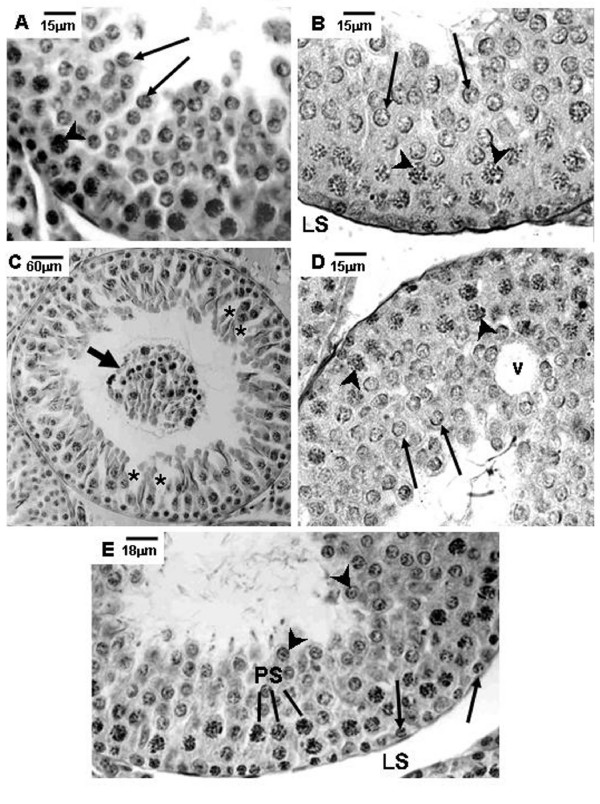
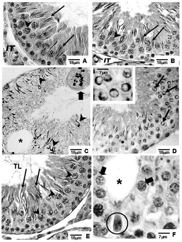
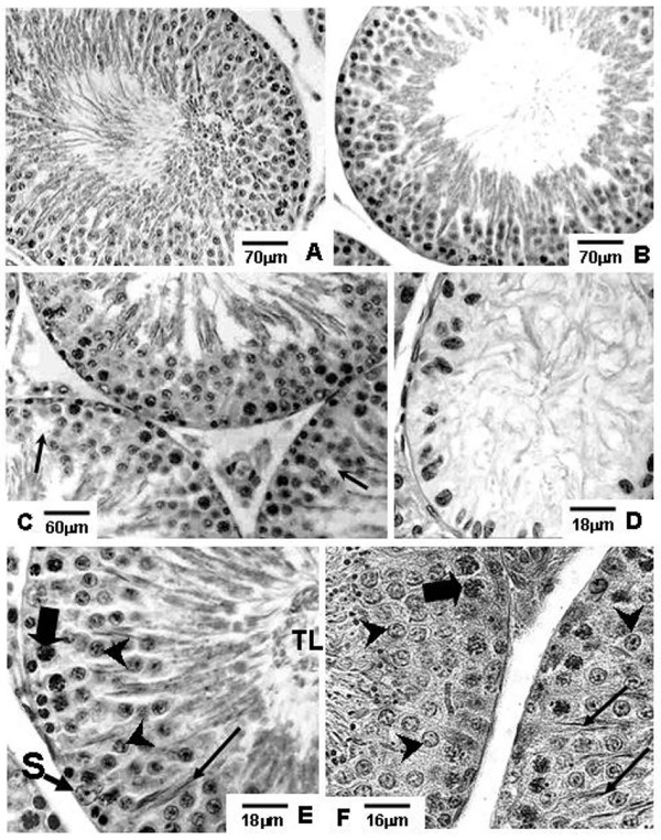
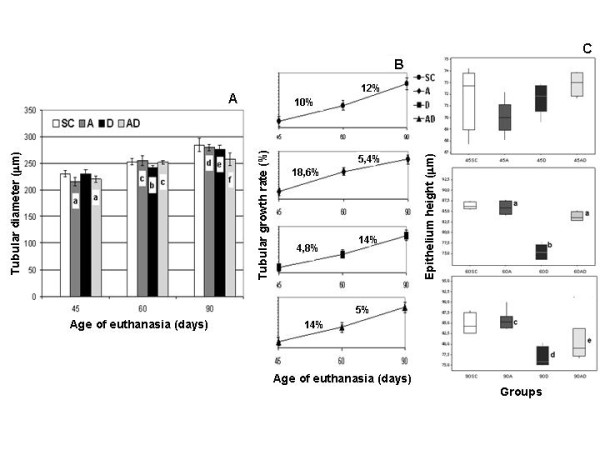
Results: The rats killed 30 and 60 days after doxorubicin treatment showed diminution of seminiferous epithelium height and reduction on the frequency of tubular sections containing at least one type of differentiated spermatogonia; reduction of sperm concentration and motility and an increase of sperm anomalous forms where observed in doxorubicin-treated animals. All these parameters were improved in the Amifostine/Doxorubicin group only when compared to Doxorubicin group. Such reduction, however, still remained below the values obtained from the Sham Control group. Nevertheless, the reproductive competence of doxorubicin-treated rats was not improved by amifostine pre-administration.
Conclusions: These results suggest that amifostine promotes a significant reduction of the doxorubicin long-term side effects on the seminiferous epithelium of prepubertal rats, which is reflected in the epidydimal fluid parameters in the adult phase. However, fertility status results suggest that such protection may not be effective against sperm DNA content damage. Further investigation of sperm DNA integrity must be carried out using amifostine and doxorubicin-treated experimental models.
Figures
Similar articles
-
Amifostine-doxorubicin association causes long-term prepubertal spermatogonia DNA damage and early developmental arrest.Hum Reprod. 2012 Aug;27(8):2457-66. doi: 10.1093/humrep/des159. Epub 2012 May 15. Hum Reprod. 2012. PMID: 22593430
-
Amifostine protective effect on cisplatin-treated rat testis.Anat Rec (Hoboken). 2008 Jul;291(7):797-808. doi: 10.1002/ar.20693. Anat Rec (Hoboken). 2008. PMID: 18543292
-
Amifostine protects against early but not late toxic effects of doxorubicin in infant rats.Cancer Res. 2001 Sep 1;61(17):6423-7. Cancer Res. 2001. PMID: 11522636
-
Cytoprotective agents for anthracyclines.Semin Oncol. 1996 Aug;23(4 Suppl 8):23-34. Semin Oncol. 1996. PMID: 8783663 Review.
-
The potential of amifostine: from cytoprotectant to therapeutic agent.Haematologica. 1999 Nov;84(11):1035-42. Haematologica. 1999. PMID: 10553165 Review.
Cited by
-
Protective Effects of Dioscin Against Doxorubicin-Induced Hepatotoxicity Via Regulation of Sirt1/FOXO1/NF-κb Signal.Front Pharmacol. 2019 Sep 13;10:1030. doi: 10.3389/fphar.2019.01030. eCollection 2019. Front Pharmacol. 2019. PMID: 31572199 Free PMC article.
-
Proanthocyanidins produce significant attenuation of doxorubicin-induced mutagenicity via suppression of oxidative stress.Oxid Med Cell Longev. 2010 Nov-Dec;3(6):404-13. doi: 10.4161/oxim.3.6.14418. Epub 2010 Nov 1. Oxid Med Cell Longev. 2010. PMID: 21311213 Free PMC article.
-
The attenuation of doxorubicin-induced testicular toxicity with improved testicular histoarchitecture of mice by the bioactive compounds in Solanum anomalum leaves: Experimental and computational studies.Toxicol Rep. 2024 Nov 21;13:101827. doi: 10.1016/j.toxrep.2024.101827. eCollection 2024 Dec. Toxicol Rep. 2024. PMID: 39649379 Free PMC article.
-
The influence of amifostine administration prior to cyclophosphamide on in vitro maturation of mouse oocytes.J Assist Reprod Genet. 2013 Jul;30(7):939-44. doi: 10.1007/s10815-013-0035-9. Epub 2013 Jul 5. J Assist Reprod Genet. 2013. PMID: 23828370 Free PMC article.
-
Stem cell-based therapies for fertility preservation in males: Current status and future prospects.World J Stem Cells. 2020 Oct 26;12(10):1097-1112. doi: 10.4252/wjsc.v12.i10.1097. World J Stem Cells. 2020. PMID: 33178394 Free PMC article. Review.
References
-
- Humpl T, Shramm P, Gutjahr P. Male fertility in long term survivors of childhood ALL. Arch Androl. 1999;43:123–129. - PubMed
Publication types
MeSH terms
Substances
LinkOut - more resources
Full Text Sources
Medical
