. 2010 Jan 11:7:4.
doi: 10.1186/1743-422X-7-4.

Classic swine fever virus NS2 protein leads to the induction of cell cycle arrest at S-phase and endoplasmic reticulum stress

Affiliations

Classic swine fever virus NS2 protein leads to the induction of cell cycle arrest at S-phase and endoplasmic reticulum stress

Qing-hai Tang et al. Virol J. .

Abstract

Background: Classical swine fever (CSF) caused by virulent strains of Classical swine fever virus (CSFV) is a haemorrhagic disease of pigs, characterized by disseminated intravascular coagulation, thrombocytopoenia and immunosuppression, and the swine endothelial vascular cell is one of the CSFV target cells. In this report, we investigated the previously unknown subcellular localization and function of CSFV NS2 protein by examining its effects on cell growth and cell cycle progression.

Results: Stable swine umbilical vein endothelial cell line (SUVEC) expressing CSFV NS2 were established and showed that the protein localized to the endoplasmic reticulum (ER). Cellular analysis revealed that replication of NS2-expressing cell lines was inhibited by 20-30% due to cell cycle arrest at S-phase. The NS2 protein also induced ER stress and activated the nuclear transcription factor kappa B (NF-kappaB). A significant increase in cyclin A transcriptional levels was observed in NS2-expressing cells but was accompanied by a concomitant increase in the proteasomal degradation of cyclin A protein. Therefore, the induction of cell cycle arrest at S-phase by CSFV NS2 protein is associated with increased turnover of cyclin A protein rather than the down-regulation of cyclin A transcription.

Conclusions: All the data suggest that CSFV NS2 protein modulate the cellular growth and cell cycle progression through inducing the S-phase arrest and provide a cellular environment that is advantageous for viral replication. These findings provide novel information on the function of the poorly characterized CSFV NS2 protein.


Figures
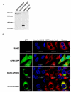
Figure 1
Figure 1
Detection and subcellular localization of the GFP-NS2 and NS2-GFP fusion protein expressed in SUVEC. (A) Western immunoblot using anti-GFP antibody of cellular proteins isolated from various cell lines. (B) Confocal microscopy images of SUVEC cells. All the cell lines were stained by Hoechst33342 and ER-Tracker™ Red. Merged images show co-localization of GFP, GFP-NS2 and NS2-GFP in the ER. Bar = 20 μm for all the figures.
Figure 2
Figure 2
Cell proliferation assays of stable NS2-expressing cell lines. The MTS assay was used to measure proliferation of 4 × 10³ cells from SUVEC cell lines over time. Each data set represents the mean ± S.D. of six replicates.
Figure 3
Figure 3
The analysis of the stages of cell division of expressing CSFV NS2 protein by flow cytometry. (A) Histograms from flow cytometry data for propidium iodide staining. (B) Analysis of the percentage of cells in each phase of the cell cycle from flow cytometry data.
Figure 4
Figure 4
The effect of CSFV NS2 expression on the porcine cyclin A protein level. (A) The level of cyclin A expression was determined by western blot with the anti-porcine cyclin A rabbit polyclonal antiserum in all cell lines treated or untreated with MG132. (B) The expression of NS2 from the same samples with and without the treatment of MG132 was detected by using anti-GFP mAb.
Figure 5
Figure 5
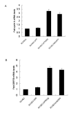
The effect of CSFV NS2 expression on porcine cyclin A and GRP78 transcription in cultured SUVEC cells. Total RNA was extracted from cells expressing either GFP alone, GFP-NS2 fusion, NS2-GFP fusion or untransfected cells. Real-time RT-PCR analysis of (A) cyclin A and (B) GRP78 mRNA levels were normalized to the corresponding CT value for porcine β-actin mRNA. The basal expression level in untransfected controls was assigned a value of 1 for each experiment.
Figure 6
Figure 6
The effect of CSFV NS2 expression on NF-κB p50 DNA-binding activity in SUVEC cell lines. NF-κB p50 activation was determined using the TransAM assay. The experiment was repeated three times and the figure shows a representative experiment.

Similar articles

Cited by

References

    1. Dreier S, Zimmermann B, Moennig V, Greiser-Wilke I. A sequence database allowing automated genotyping of Classical swine fever virus isolates. J Virol Methods. 2007;140:95–99. doi: 10.1016/j.jviromet.2006.11.013. - DOI - PubMed
    1. Harada T, Tautz N, Thiel HJ. E2-p7 region of the bovine viral diarrhea virus polyprotein: processing and functional studies. J Virol. 2000;74:9498–9506. doi: 10.1128/JVI.74.20.9498-9506.2000. - DOI - PMC - PubMed
    1. Agapov EV, Murray CL, Frolov I, Qu L, Myers TM, Rice CM. Uncleaved NS2-3 is required for production of infectious bovine viral diarrhea virus. J Virol. 2004;78:2414–2425. doi: 10.1128/JVI.78.5.2414-2425.2004. - DOI - PMC - PubMed
    1. Elbers K, Tautz N, Becher P, Stoll D, Rumenapf T, Thiel HJ. Processing in the pestivirus E2-NS2 region: identification of proteins p7 and E2p7. J Virol. 1996;70:4131–4135. - PMC - PubMed
    1. Lackner T, Thiel HJ, Tautz N. Dissection of a viral autoprotease elucidates a function of a cellular chaperone in proteolysis. Proc Natl Acad Sci USA. 2006;103:1510–1515. doi: 10.1073/pnas.0508247103. - DOI - PMC - PubMed

Publication types

LinkOut - more resources