. 2010 Jan 11:7:2.
doi: 10.1186/1742-2094-7-2.

Norepinephrine enhances the LPS-induced expression of COX-2 and secretion of PGE2 in primary rat microglia

Affiliations

Norepinephrine enhances the LPS-induced expression of COX-2 and secretion of PGE2 in primary rat microglia

Johannes C M Schlachetzki et al. J Neuroinflammation. .

Abstract

Background: Recent studies suggest an important role for neurotransmitters as modulators of inflammation. Neuroinflammatory mediators such as cytokines and molecules of the arachidonic acid pathway are generated and released by microglia. The monoamine norepinephrine reduces the production of cytokines by activated microglia in vitro. However, little is known about the effects of norepinephrine on prostanoid synthesis. In the present study, we investigate the role of norepinephrine on cyclooxygenase- (COX-)2 expression/synthesis and prostaglandin (PG)E2 production in rat primary microglia.

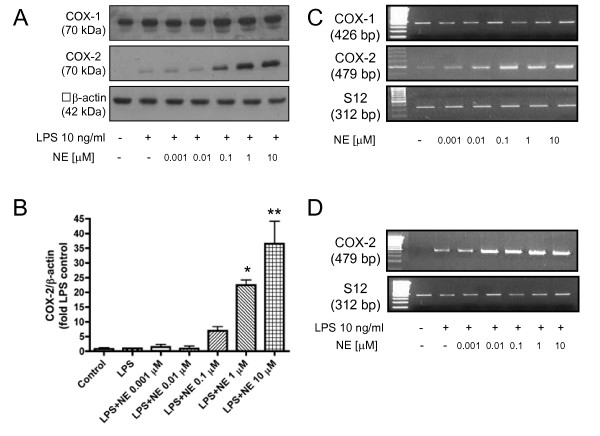
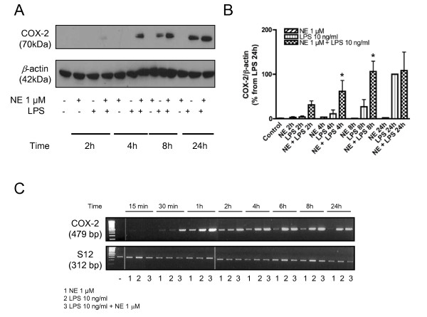
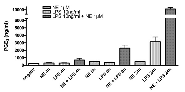
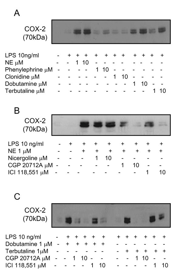
Results: Interestingly, norepinephrine increased COX-2 mRNA, but not protein expression. Norepinephrine strongly enhanced COX-2 expression and PGE2 production induced by lipopolysaccharide (LPS). This effect is likely to be mediated by beta-adrenoreceptors, since beta-, but not alpha-adrenoreceptor agonists produced similar results. Furthermore, beta-adrenoreceptor antagonists blocked the enhancement of COX-2 levels induced by norepinephrine and beta-adrenoreceptor agonists.

Conclusions: Considering that PGE2 displays different roles in neuroinflammatory and neurodegenerative disorders, norepinephrine may play an important function in the modulation of these processes in pathophysiological conditions.


Figures

Figure 1
Figure 1
Different concentrations of norepinephrine enhance the synthesis of COX-2 protein (A, B) and mRNA (C) in LPS-stimulated primary neonatal microglial cells. Microglial cells were stimulated with different concentrations of norepinephrine (0.001 μM to 10 μM) and LPS (10 ng/ml) for 4 hours. (A) A representative western blot against β-actin (42 kDa) and COX-2 (70 kDa) is shown here. (B) Quantitative densitometric analysis of COX-2 protein expression normalized to β-actin control (n = 3). *P < 0.05, **P < 0.001 with respect to LPS control. (C, D) Semi-quantitative PCR analysis of the effect of different concentrations of norepinephrine alone (C) and of norepinephrine plus LPS on COX-2 and COX-1 mRNA expression (D). [NE = norepinephrine].
Figure 2
Figure 2
Time course of treatment with norepinephrine 1 μM alone, LPS 10 ng/ml alone, or a combination of norepinephrine 1 μM plus LPS 10 ng/ml on the expression of COX-2 protein and mRNA (A-C). (A, B) Microglial cells were stimulated for 4, 8, and 24 hours. This was followed by western blot analysis against β-actin (42 kDa) and COX-2 (70 kDa). A representative western blot is shown here (A). Quantitative densitometric analysis of COX-2 normalized to β-actin loading control. *P < 0.05 with respect to LPS control. (C) Semi-quantitative analysis of the effect of the different treatment groups on COX-2 mRNA expression at different time points (15 min to 24 hours). [NE = norepinephrine].
Figure 3
Figure 3
Effect of norepinephrine on PGE2 concentrations at different time points (0 h, 4 h, 8 h, 24 h) was measured in the supernatants of LPS-stimulated primary microglial cell cultures using EIA (n = 6). Data are depicted as means ± standard error of mean. Statistical analysis was done using ANOVA with post hoc Newman-Keuls for the 4 different time points (*P < 0.001). [NE = norepinephrine].
Figure 4
Figure 4
Effect of different α- and β-adrenergic agonists and antagonists on COX-2 protein expression. Microglial cells were stimulated with LPS 10 ng/ml and different adrenergic agonists/antagonists for 4 hours, followed by western blot analysis for COX-2 protein expression (70 kDa). (A) LPS-stimulated microglial cells were treated with different α- and β-adrenoreceptor agonists at two different concentrations (1 and 10 μM): α1-agonist phenylephrine; α2-agonist clonidine; β12-agonist dobutamine; β2-agonist terbutaline. (B) LPS-stimulated microglial cells were treated with different α- and β-adrenoreceptor antagonists at two different concentrations (1 and 10 μM): α12-antagonist nicergoline; β1-antagonist CGP 20712A; β2-antagonist ICI118, 551. (C) LPS-stimulated microglial cells were treated both with dobutamine or terbutaline and with the two different β-adrenoreceptor antagonists ICI 118,551 or CGP 20712A. [NE = norepinephrine].

References

    1. Nimmerjahn A, Kirchhoff F, Helmchen F. Resting microglial cells are highly dynamic surveillants of brain parenchyma in vivo. Science. 2005;308:1314–1318. doi: 10.1126/science.1110647. - DOI - PubMed
    1. Davalos D, Grutzendler J, Yang G, Kim JV, Zuo Y, Jung S, Littman DR, Dustin ML, Gan WB. ATP mediates rapid microglial response to local brain injury in vivo. Nat Neurosci. 2005;8:752–758. doi: 10.1038/nn1472. - DOI - PubMed
    1. Hanisch UK, Kettenmann H. Microglia: active sensor and versatile effector cells in the normal and pathologic brain. Nat Neurosci. 2007;10:1387–1394. doi: 10.1038/nn1997. - DOI - PubMed
    1. Block ML, Zecca L, Hong JS. Microglia-mediated neurotoxicity: uncovering the molecular mechanisms. Nat Rev Neurosci. 2007;8:57–69. doi: 10.1038/nrn2038. - DOI - PubMed
    1. Wright SD, Ramos RA, Tobias PS, Ulevitch RJ, Mathison JC. CD14, a receptor for complexes of lipopolysaccharide (LPS) and LPS binding protein. Science. 1990;249:1431–1433. doi: 10.1126/science.1698311. - DOI - PubMed

Publication types

MeSH terms