. 2010 Jan 11:10:6.
doi: 10.1186/1471-2148-10-6.

Molecular and cellular evidence for biased mitotic gene conversion in hybrid scallop

Affiliations

Molecular and cellular evidence for biased mitotic gene conversion in hybrid scallop

Shi Wang et al. BMC Evol Biol. .

Abstract

Background: Concerted evolution has been believed to account for homogenization of genes within multigene families. However, the exact mechanisms involved in the homogenization have been under debate. Use of interspecific hybrid system allows detection of greater level of sequence variation, and therefore, provide advantage for tracing the sequence changes. In this work, we have used an interspecific hybrid system of scallop to study the sequence homogenization processes of rRNA genes.

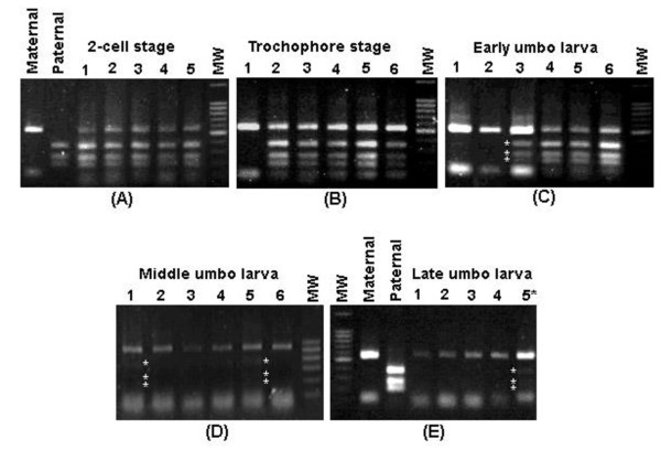
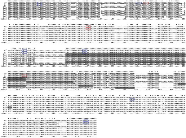
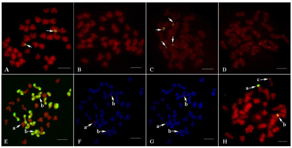
Results: Through the use of a hybrid scallop system (Chlamys farreri female symbol x Argopecten irradians male symbol), here we provide solid molecular and cellular evidence for homogenization of the rDNA sequences into maternal genotypes. The ITS regions of the rDNA of the two scallop species exhibit distinct sequences and thereby restriction fragment length polymorphism (RFLP) patterns, and such a difference was exploited to follow the parental ITS contributions in the F1 hybrid during early development using PCR-RFLP. The representation of the paternal ITS decreased gradually in the hybrid during the development of the hybrid, and almost diminished at the 14th day after fertilization while the representation of the maternal ITS gradually increased. Chromosomal-specific fluorescence in situ hybridization (FISH) analysis in the hybrid revealed the presence of maternal ITS sequences on the paternal ITS-bearing chromosomes, but not vice versa. Sequence analysis of the ITS region in the hybrid not only confirmed the maternally biased conversion, but also allowed the detection of six recombinant variants in the hybrid involving short recombination regions, suggesting that site-specific recombination may be involved in the maternally biased gene conversion.

Conclusion: Taken together, these molecular and cellular evidences support rapid concerted gene evolution via maternally biased gene conversion. As such a process would lead to the expression of only one parental genotype, and have the opportunities to generate recombinant intermediates; this work may also have implications in novel hybrid zone alleles and genetic imprinting, as well as in concerted gene evolution. In the course of evolution, many species may have evolved involving some levels of hybridization, intra- or interspecific, the sex-biased sequence homogenization could have led to a greater role of one sex than the other in some species.


Figures

Figure 1
Figure 1
PCR-RFLP analysis of make-up of the ITS region in the hybrid with restriction enzyme Hae III at early development. Panel A exhibits PCR-RFLP analysis at the 2-cell stage (about 1 hour after fertilization) with samples from maternal parent C. farreri (Maternal), paternal parent A. irradians (Paternal), and samples from five random selected hybrid individuals (1 through 5), and molecular weight standard (MW). Panel B exhibits PCR-RFLP analysis at the trochophore stage (about 20 hour after fertilization) with samples from six random selected hybrid samples (1 through 6), and molecular weight standard (MW). Panel C exhibits PCR-RFLP analysis at the early umbo larva stage (about 4 days after fertilization) with samples from six random selected hybrid samples (1 through 6), and molecular weight standard (MW). Panel D exhibits PCR-RFLP analysis at the middle umbo larva stage (about 10 days after fertilization) with samples from six random selected hybrid individuals (1 through 6), and molecular weight standard (MW). Panel E exhibits PCR-RFLP analysis at the late umbo larva stage (about 14 days after fertilization) with samples from maternal parent (Maternal), paternal parent (Paternal), and samples from four random selected hybrid individuals (1 through 4) and a sample from mixed samples of multiple individuals (5*), and molecular weight standard (MW). Molecular weight standards were 100 bp DNA ladder.
Figure 2
Figure 2
Alignment of parental and 6 recombinant variants' sequences. Cf: C. farreri; Ai: A. irradians; RV1-6: recombinant variants 1-6. Potential recombinant regions in recombinant variants are shown in black background. Asterisks show identical bases; dashes indicate alignment gaps. (ITS1: 34-350 bp; 5.8S: 351-506 bp; ITS2: 507-805 bp). Restriction sites for Mbo I GATC are indicated by a blue box while restriction sites for Hae III GGCC are indicated by a red box.
Figure 3
Figure 3
Schematic presentation of the recombinant variants of the interspecific hybrid showing the origins of the sequences in the ITS region. Open bar, sequences from maternal parent; sketched bar, sequences from paternal parent. Restriction sites for Mbo I are indicated by blue arrows while restriction sites for Hae III are indicated by red arrows.
Figure 4
Figure 4
Chromosomal locations of the parental ITSs in the hybrid. (A) FISH in C. farreri with probe from C. farreri (signals are shown by arrows); (B) FISH in C. farreri with probe from A. irradians (no signal is detected); (C) FISH in A. irradians with probe from A. irradians (signals are shown by arrows); (D) FISH in A. irradians with probe from C. farreri (no signal is detected); (E) GISH in the hybrid with genome probe from A. irradians (a shows the ITS-bearing chromosome of C. farreri; b show the ITS-bearing chromosomes of A. irradians; metaphases in E, F and G are same); (F) FISH in the hybrid with probe from C. farreri (signals are detected at a and b); (G) FISH in the hybrid with probe from A. irradians (no signal is detected at a but weak signals are detected at b); (H) FISH in the hybrid with probe from C. farreri (a shows the ITS-bearing chromosome of C. farreri; b, c show the ITS-bearing chromosomes of A. irradians; signal intensity at b is higher than that at c). Scale bar represents 5 μm.

References

    1. Nei M, Rooney AP. Concerted and birth-and-death evolution of multigene families. Annu Rev Genet. 2005;39:121–152. doi: 10.1146/annurev.genet.39.073003.112240. - DOI - PMC - PubMed
    1. Dover G. Molecular drive: a cohesive mode of species evolution. Nature. 1982;299(5879):111–117. doi: 10.1038/299111a0. - DOI - PubMed
    1. Eickbush TH, Eickbush DG. Finely orchestrated movements: evolution of the ribosomal RNA genes. Genetics. 2007;175:477–85. doi: 10.1534/genetics.107.071399. - DOI - PMC - PubMed
    1. Gonzalez IL, Sylvester JE. Human rDNA: evolutionary patterns within the genes and tandem arrays derived from multiple chromosomes. Genomics. 2001;73(3):255–263. doi: 10.1006/geno.2001.6540. - DOI - PubMed
    1. Hughes KW, Petersen RH. Apparent recombination or gene conversion in the ribosomal ITS region of a Flammulina (Fungi, Agaricales) hybrid. Mol Biol Evol. 2001;18(1):94–96. - PubMed

Publication types

Substances