Review
. 2009 Dec 24;139(7):1229-41.
doi: 10.1016/j.cell.2009.12.008.

Metabolism, cell surface organization, and disease

Affiliations
Review

Metabolism, cell surface organization, and disease

James W Dennis et al. Cell. .

Abstract

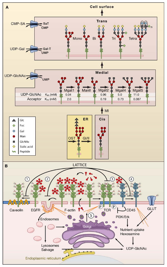
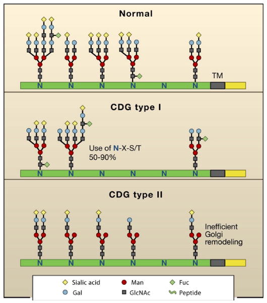
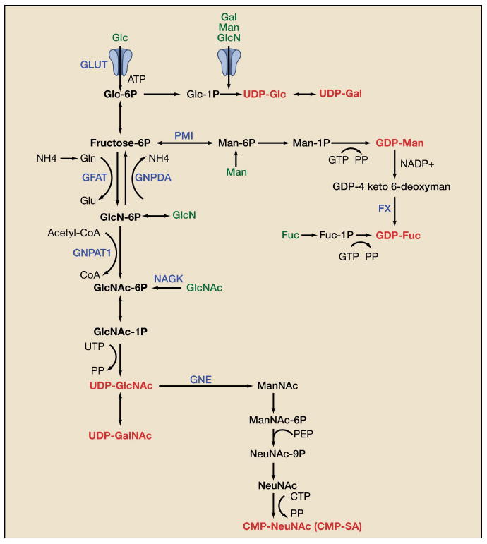
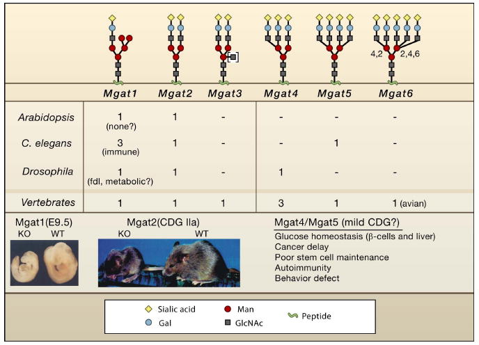
Genetic information flows from DNA to macromolecular structures-the dominant force in the molecular organization of life. However, recent work suggests that metabolite availability to the hexosamine and Golgi N-glycosylation pathways exerts control over the assembly of macromolecular complexes on the cell surface and, in this capacity, acts upstream of signaling and gene expression. The structure and number of N-glycans per protein molecule cooperate to regulate lectin binding and thereby the distribution of glycoproteins at the cell surface. Congenital disorders of glycosylation provide insight as extreme hypomorphisms, whereas milder deficiencies may encompass many common chronic conditions, including autoimmunity, metabolic syndrome, and aging.


Figures

Figure 1
Figure 1. N-glycan branching pathways and the galectin lattice
(A) N-glycan branching pathway. Oligosaccharyltransterase (OT) utilizes the pre-assembled donor Glc3Man9GlcNAc2-pp-dolichol to transfer the glycan to N-X-S/T motifs on glycoproteins in the endoplasmic reticulum (ER). Glycoproteins transit from the ER to cis, medial, and trans Golgi en route to the cell surface. The N-acetylglucosaminyltransferases enzymes, designated by their gene names (Mgat1, Mgat2, Mgat4, Mgat5) generate branched N-glycans that display a range of affinities for galectins. The Km values for Mgat1, Mgat2, Mgat4 and Mgat5 are indicated as measured in vitro for UDP-GlcNAc and acceptor glycoproteins. (B) Dynamics of the galectin lattice. The glycocalyx is the thick carbohydrate layer surrounding the cell. Glycan structures generated in the Golgi differ in affinities for galectins. Galectins cross-link glycoprotein receptors and oppose (1) loss of epidermal growth factor receptors (EGFR) to Caveolin 1-positive microdomains, (2) coated-pit endocytosis, (3) precocious clustering of receptors, and (4) F-actin-mediated entry of T cell receptor (TCR) into and exit of CD45 from ganglioside GM1-positive microdomains (blue). (5) Nutrient supply and growth signaling increase membrane remodeling, regulate metabolite flux through the hexosamine pathway to UDP-GlcNAc and N-glycan branching (Golgi) on receptors to promote surface retention by the galectin lattice.
Figure 2
Figure 2. Congenital Disorders of Glycosylation and glycoforms
(A) Glycoprotein isoforms or glycofoms differ based on N-glycan structures present at each N-X-S/T site (not all sites are occupied). CDG type I alters glycoform distribution due to deficiencies in Glc3Man9GlcNAc2-pp-dolichol biosynthesis leading to incomplete N-X-S/T site occupancy. CDG type II are deficiencies in Golgi remodeling or glycoprotein trafficking.
Figure 3
Figure 3. Biosynthesis of Sugar-nucleotides
De novo biosynthesis of UDP-GlcNAc is dependent on glucose, glutamine, acetyl-CoA and uridine. UDP-GlcNAc is also the precursor for UDP-GalNAc and CMP-SA biosynthesis. Gal, Man, and GlcN salvage/import (green) are facilitated by the hexose transporters (GLUTs), whereas GlcNAc and Fuc are taken up by bulk endocytosis. The sugar nucleotides are in red. Genes in blue are discussed in the text. Metabolite abbreviations: glucose (Glc), galactose (Gal), mannose (Man), glucosamine (GlcN), fucose (Fuc), N-acetylgalactosamine (GalNAc), N-acetylglucosamine (GlcNAc), N-acetylneuraminic acid (NeuNAc), glutamine (Gln).
Figure 4
Figure 4. Mgat deficiencies
Listed are the number of genes encoding Mgat activities corresponding to the branching pattern shown above. C. elegans triple Mgat1 mutant worms develop normally, but display altered sensitivities to pathogenic bacteria (Shi et al., 2006). Mutation of Mgat1 in Drosophila leads to fused lobes in the brain, and loss of motility in adult flies, possibly reflecting both developmental and metabolic deficiencies (Sarkar et al., 2006). A fused lobes (fdl) phenotype is also observed for loss of β-hexosaminidase, the enzyme that specifically removes GlcNAc added by Mgat1 (Leonard et al., 2006). It is possible that a precise level of Mgat1 product regulates a morphogen gradient that defines the boundary between lobes. Mgat1 deficient embryos are growth impaired and display defects in neural tube closure (Ioffe and Stanley, 1994; Metzler et al., 1994). Mice lacking Mgat2 are small and display severe defects in multiple organs, which is comparable to human CDG-IIa. Reproduced by permission of Oxford University Press: Wang et al. (2001) Glycobiology, 11 (12), 1051-1070. The product of Mgat3 shown in brackets can be found in any of the structures shown, but its presence blocks further branching.

Similar articles

Cited by

References

    1. Ahmad N, Gabius HJ, Andre S, Kaltner H, Sabesan S, Roy R, Liu B, Macaluso F, Brewer CF. Galectin-3 precipitates as a pentamer with synthetic multivalent carbohydrates and forms heterogeneous cross-linked complexes. Journal of Biological Chemistry. 2003;279:10841–10847. - PubMed
    1. Alegre ML, Frauwirth KA, Thompson CB. T-cell regulation by CD28 and CTLA-4. Nat Rev Immunol. 2001;1:220–228. - PubMed
    1. Amano M, Galvan M, He J, Baum LG. The ST6Gal I sialyltransferase selectively modifies N-glycans on CD45 to negatively regulate galectin-1-induced CD45 clustering, phosphatase modulation, and T cell death. J Biol Chem. 2003;278:7469–7475. - PubMed
    1. Anjos S, Nguyen A, Ounissi-Benkalha H, Tessier MC, Polychronakos C. A common autoimmunity predisposing signal peptide variant of the cytotoxic T-lymphocyte antigen 4 results in inefficient glycosylation of the susceptibility allele. J Biol Chem. 2002;277:46478–46486. - PubMed
    1. Aoki M, Lin CL, Rothstein JD, Geller BA, Hosler BA, Munsat TL, Horvitz HR, Brown RH., Jr Mutations in the glutamate transporter EAAT2 gene do not cause abnormal EAAT2 transcripts in amyotrophic lateral sclerosis. Ann Neurol. 1998;43:645–653. - PubMed

Publication types