. 2009 Dec 24;139(7):1255-67.
doi: 10.1016/j.cell.2009.12.018.

A physical and regulatory map of host-influenza interactions reveals pathways in H1N1 infection

Affiliations

A physical and regulatory map of host-influenza interactions reveals pathways in H1N1 infection

Sagi D Shapira et al. Cell. .

Abstract

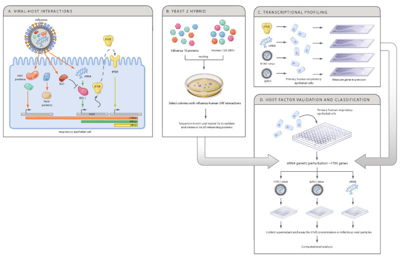
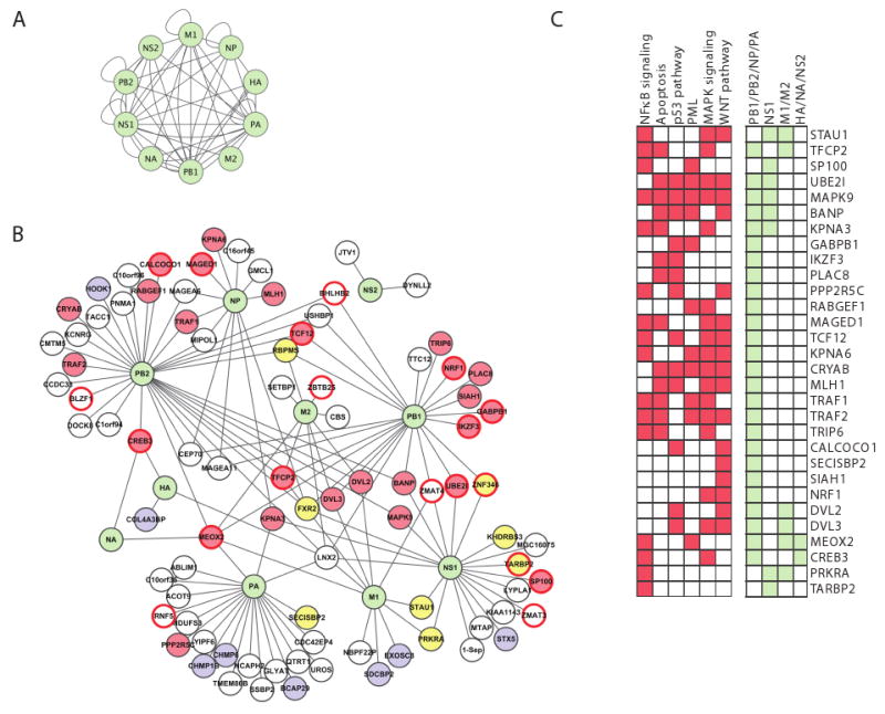
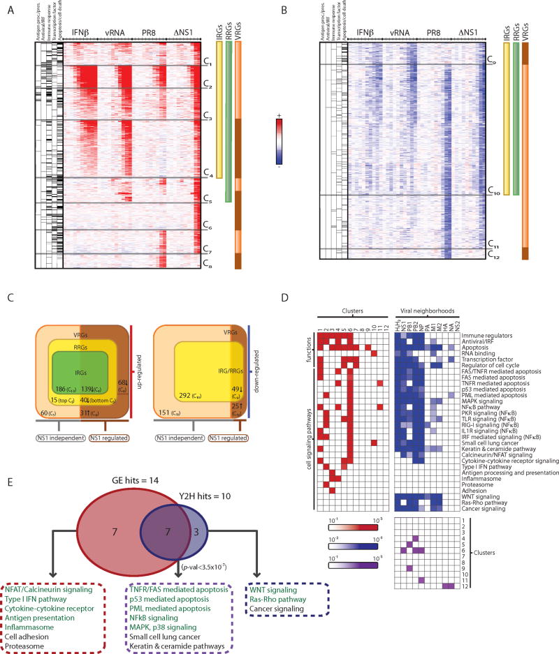
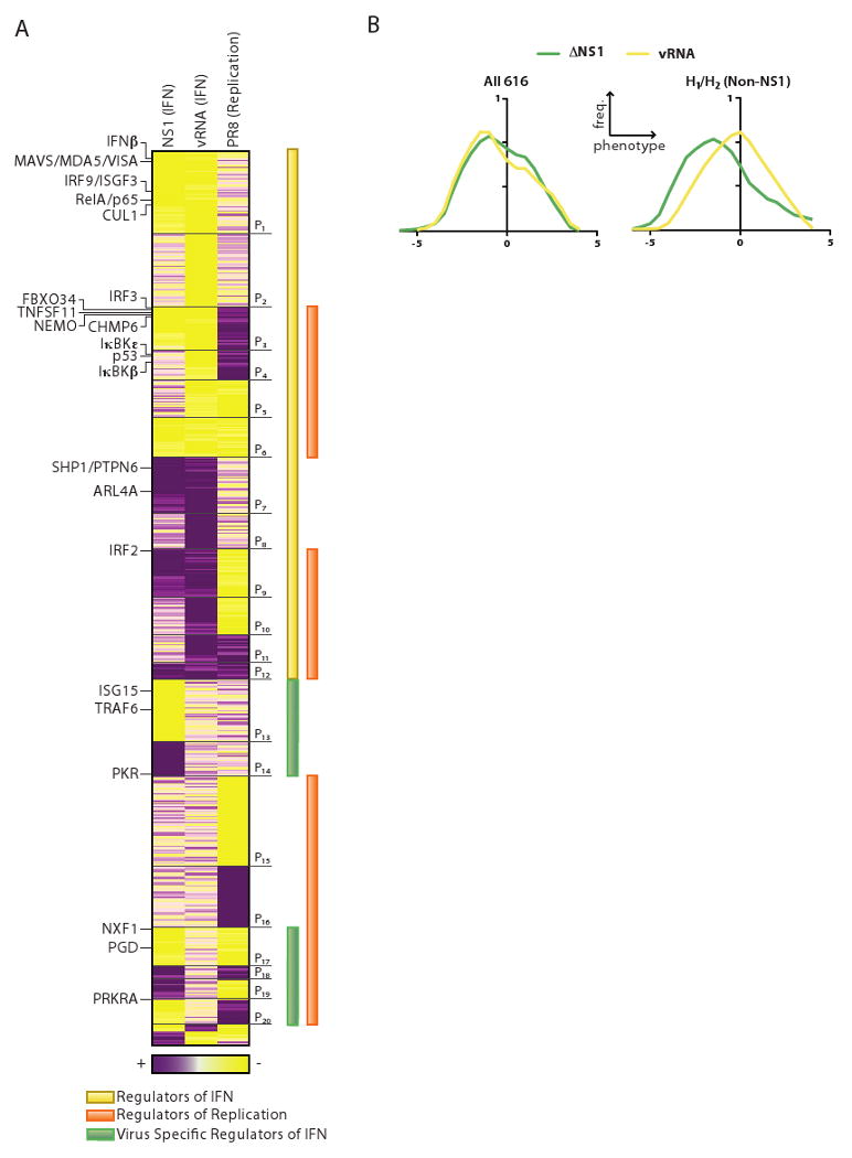
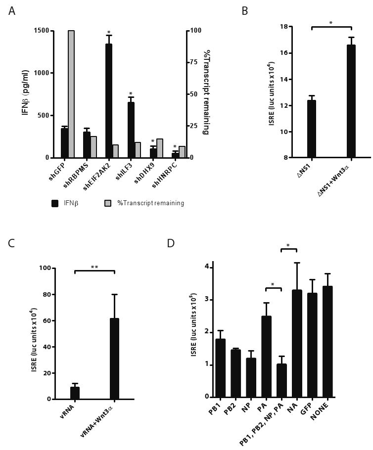
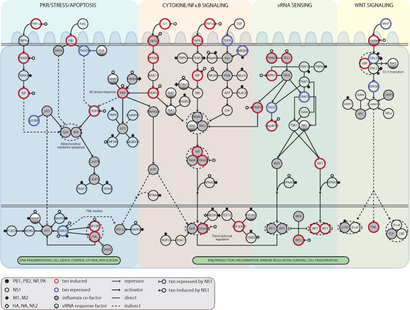
During the course of a viral infection, viral proteins interact with an array of host proteins and pathways. Here, we present a systematic strategy to elucidate the dynamic interactions between H1N1 influenza and its human host. A combination of yeast two-hybrid analysis and genome-wide expression profiling implicated hundreds of human factors in mediating viral-host interactions. These factors were then examined functionally through depletion analyses in primary lung cells. The resulting data point to potential roles for some unanticipated host and viral proteins in viral infection and the host response, including a network of RNA-binding proteins, components of WNT signaling, and viral polymerase subunits. This multilayered approach provides a comprehensive and unbiased physical and regulatory model of influenza-host interactions and demonstrates a general strategy for uncovering complex host-pathogen relationships.


Figures

Figure 1
Figure 1. Integrative strategy to generate a physical, regulatory and functional map of influenza-host interactions
(A) When influenza infects host cells, viral components, including viral RNA (vRNA) and viral proteins interact with host proteins to induce changes in host gene expression or cellular functions. The RIG-I sensor detects virus-derived RNA and regulates host gene expression, including IFNβ, which in turn activates an anti-viral program through the interferon receptor (IFNR). We distinguish viral regulated genes (VRGs, orange), affected by infection, RNA regulated genes (RRGs, green), affected directly by vRNA, and interferon regulated genes (IRGs, yellow), affected directly by interferon treatment. NS1 is shown to inhibit the RIG-I response. (B-D) A genomic strategy to deconstruct influenza-host interactions. Host proteins that physically interact with each of the 10 viral proteins are identified using a systematic yeast-2-hybrid approach (B), and arrays are used to define the transcriptional responses of primary human bronchial epithelial cells (HBECs) to components of the virus and to virus infection (C). The physical and transcriptional maps were used to computationally predict human factors and pathways that affect the viral life cycle or host response. We tested these predictions by perturbing each gene and measuring the effect on IFN production and viral replication in primary HBECs (D).
Figure 2
Figure 2. A map of viral-human protein interactions identifies a dense interconnected network, coupled to key cellular signaling pathways. (A) Viral-viral interactions
Green nodes, viral proteins; edges, direct physical interactions observed in a Y2H assay. (B) Influenza-human interactions. 134 interactions (edges) connect the ten influenza proteins (green nodes) to 87 ‘H1’ human proteins. Yellow fill, RNA-binding proteins; blue fill, protein transport; red border, transcription factors; red fill, 30 proteins that play a role in four major signaling pathways (NFκB, apoptosis, MAPK and WNT signaling); white fill, proteins with other functions. (C) 30 host interactor proteins (H1) are shown with their membership in specific pathways (red) or direct interactions with influenza proteins (light green). H1 proteins are either known components or established direct interactors with components of these pathways (see Experimental Procedures). Many of the 30 proteins are involved in multiple signaling pathways, and interact with polymerase subunits. Description of influenza A proteins: PB1, PB2, PA form the viral polymerase; NP complexes with viral RNA and is required for viral polymerase activity on RNA; HA and NA are membrane proteins involved in the fusion and release of viral particles; M1 is a matrix protein involved in export and assembly of RNA and viral particles; M2 modulates fusion through its ion channel activity; NS1 is a non-structural protein that regulates host pathways; NS2 is involved in RNA export.
Figure 3
Figure 3. Functional decomposition of transcriptional responses to influenza infection identifies viral-specific gene regulation
(A, B) Gene expression changes in HBECs in response to IFNβ (IRGs, yellow bar), vRNA (RRGs, green bar), wild type influenza (PR8) and mutant ΔNS1 virus (VRGs, orange bar; genes differentially regulated by NS1, brown bar) at 10 time points (.25, .5, 1, 1.5, 2, 4, 6, 8, 12, 18 hours, tick marks). Upregulated (A, red) and downregulated (B, blue) genes, relative to the expected level from mock-treated cells, were grouped into 12 clusters (C1-C12). Left columns denote gene membership in five major functional categories (black lines, category is enriched in cluster; grey lines, category is not enriched in cluster). (C) Venn diagrams indicate number of members in each class of regulated genes and their dependence on NS1 (bottom), within the subset of upregulated (left) and downregulated (right) genes. (D) Functional and pathway annotation of expression clusters and interaction neighborhoods. Shown are the functional categories and pathways (rows) enriched in each of the 12 expression clusters (red, left matrix) and interaction neighborhoods (H1 and H2) of each viral protein (blue, right matrix). Bottom matrix shows the significant overlaps (purple) between expression clusters (1-12, rows) and viral neighborhoods (columns). (E) Enrichment analysis identifies pathways that are over-represented in the influenza physical network (10 pathways, blue) and in transcriptional responses (14 pathways, red), with an overlap of 7 pathways enriched in both (purple, P <3.5×10⁻⁷). Pathways chosen for functional follow-up assays are colored in green.
Figure 4
Figure 4. Functional interrogation and classification of candidate genes identified through integrative analysis of influenza-human interactions
(A) Classification of phenotypes resulting from siRNA-mediated knockdown of 616 genes. Heat map shows phenotype scores corresponding to 3 functional assays (columns) performed on HBECs following transfection with siRNAs (rows). Gene phenotypes are hierarchically clustered, resulting in 20 major phenoclusters (P1-20). NS1 (IFN), assay for production of interferon following infection with ΔNS1 virus. vRNA (IFN), assay for production of interferon following transfection with viral RNA. PR8 (Replication), assay for infectious virion production following infection with PR8 virus. Yellow, positive regulator (lower IFN or virus titer). Purple, negative regulator (higher IFN or virus titer). Selected genes (also referred to in the text) are marked (left). (B) Distribution of phenotype scores for direct physical interactors and their first neighbors. Right panel: H1/H2 interactors (excluding NS1) have a significantly higher number of positive regulators of interferon production following infection with ΔNS1 (green) compared to vRNA transfection (yellow). There is no such shift in the distribution for all 616 genes shown in the phenocluster heatmap (left panel).
Figure 5
Figure 5. Functional roles of an RNA-binding protein subnetwork, the WNT pathway and the viral polymerase
(A) RNA binding proteins play a role in regulating interferon production in HBECs infected with ΔNS1 virus. HBECs were infected with lentiviral shRNAs to knockdown each of 5 candidate RNA-binding proteins. Cells were selected in puromycin for 5 days and then stimulated with ΔNS1 virus. Supernatants were collected 24 hours post-infection, and IFNβ protein levels were quantified by ELISA (black bars, left Y-axis). In the same experiment, the efficacy of knockdown by each shRNA on its target mRNA was quantified (grey bars, right Y-axis, measured by qPCR relative to GAPDH). (n=3). (B,C) WNT protein potentiates ISRE responses in epithelial cells. 293T-ISRE-luciferase reporter cells were treated with WNT3α for 24 hrs and then infected with ΔNS1 virus or transfected with vRNA for 18 hours. Luciferase reporter activity (Y-axis) was quantified in response to (B) ΔNS1 infection or (C) transfected vRNA (purified from PR8 virions). (n=6). (D) Overexpression of viral polymerase subunits or NP and their effects on ISRE-inducing activity following vRNA transfection. 293T-ISRE-luciferase reporter cells were transfected with an expression plasmid encoding each influenza polymerase subunit or with combinations of plasmids (bottom panel) and then stimulated with transfected vRNA. ISRE responses to transfected vRNA were quantified for each of PB1, PB2, NP, PA, NA and control GFP, and their combinations. Similar results were obtained when cells were infected with ΔNS1 virus (data not shown). Significant effects of overexpression were found only for PB1, PB2, NP, PB1/PB2/NP/PA vs. NA (but only two are marked for clarity). (n=6). Error bars represent the standard deviation of the replicates. *, p<0.05 (t-test).
Figure 6
Figure 6. An integrated model of the key signaling pathways modulated by influenza-host interactions
For each host component within computationally selected pathways (see text), we show its mode of regulation and its functional role: (i) direct physical contact with viral proteins (small circles and diamonds); NS1 interactions with PKR, RIG-I, TRIM25 were added manually based on previous reports; (ii) transcriptional regulation in response to influenza infection in HBECs (increase in gene expression, thick red border; decrease, thick blue border); (iii) transcriptional regulation by the NS1 protein (open circle with inhibitory edge or activating edge); (iv) role in modulation of IFN production or PR8 replication (filled, gray); or role only in the IFN response to vRNA (filled, gradient). Txn, transcriptional. Influenza co-factor indicates significant change in PR8 replication upon siRNA knockdown of that gene. vRNA response factor indicates a gene whose knockdown caused significant change only in the vRNA-induced interferon assay.

Similar articles

Cited by

References

    1. Ashburner M, Ball CA, Blake JA, Botstein D, Butler H, Cherry JM, Davis AP, Dolinski K, Dwight SS, Eppig JT, et al. Gene ontology: tool for the unification of biology. The Gene Ontology Consortium. Nat Genet. 2000;25:25–29. - PMC - PubMed
    1. Bailer SM, Haas J. Connecting viral with cellular interactomes. Curr Opin Microbiol. 2009;12:453–459. - PMC - PubMed
    1. Brander C, Walker BD. Modulation of host immune responses by clinically relevant human DNA and RNA viruses. Curr Opin Microbiol. 2000;3:379–386. - PubMed
    1. Brass AL, Dykxhoorn DM, Benita Y, Yan N, Engelman A, Xavier RJ, Lieberman J, Elledge SJ. Identification of host proteins required for HIV infection through a functional genomic screen. Science. 2008;319:921–926. - PubMed
    1. Bushman FD, Malani N, Fernandes J, D'Orso I, Cagney G, Diamond TL, Zhou H, Hazuda DJ, Espeseth AS, Konig R, et al. Host cell factors in HIV replication: meta-analysis of genome-wide studies. PLoS Pathog. 2009;5:e1000437. - PMC - PubMed

Publication types

MeSH terms

Associated data