. 2009 Dec 24;64(6):841-56.
doi: 10.1016/j.neuron.2009.11.003.

Molecular identification of rapidly adapting mechanoreceptors and their developmental dependence on ret signaling

Affiliations

Molecular identification of rapidly adapting mechanoreceptors and their developmental dependence on ret signaling

Wenqin Luo et al. Neuron. .

Abstract

In mammals, the first step in the perception of form and texture is the activation of trigeminal or dorsal root ganglion (DRG) mechanosensory neurons, which are classified as either rapidly (RA) or slowly adapting (SA) according to their rates of adaptation to sustained stimuli. The molecular identities and mechanisms of development of RA and SA mechanoreceptors are largely unknown. We found that the "early Ret(+)" DRG neurons are RA mechanoreceptors, which form Meissner corpuscles, Pacinian corpuscles, and longitudinal lanceolate endings. The central projections of these RA mechanoreceptors innervate layers III through V of the spinal cord and terminate within discrete subdomains of the dorsal column nuclei. Moreover, mice lacking Ret signaling components are devoid of Pacinian corpuscles and exhibit a dramatic disruption of RA mechanoreceptor projections to both the spinal cord and medulla. Thus, the early Ret(+) neurons are RA mechanoreceptors and Ret signaling is required for the assembly of neural circuits underlying touch perception.


Figures

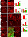
Figure 1
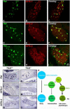
Figure 1. The early Ret+ DRG neurons express GFRα2
Double fluorescent in situ hybridization of Ret and GFRα1 (A–C), Ret and GFRα2 (D–F), and Ret and GFRα3 (G–I) at E13.5 (n=3, and 6 to 8 sections were examined for each animal, lower lumbar DRGs). J–O: In situ hybridization of Ret (37 ± 6 vs. 7 ± 3), GFRα1 (29 ± 3 vs. 0), and GFRα2 (10 ± 3 vs. 9 ± 1) in control and TrkA null DRGs at E13.5 (control vs. mutant, mean ± SEM positive neurons/section, n=3 animals per genotype, and 6 to 8 sections were examined and quantified for each animal, lower lumbar DRGs). P. Illustration of the three populations of Ret+ DRG neurons based on their developmental origin and temporal patterns of expression of Ret and GFRα co-receptors.
Figure 2
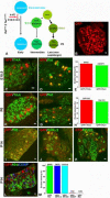
Figure 2. Genetic Labeling of the early Ret+ DRG neurons
A. Outline of the chemical-genetic strategy for labeling the early Ret+ neurons. RetERT2 and Tauf(mGFP) mice were crossed, and timed pregnant mothers were gavaged with 1.0 to 1.5 mg 4-HT per day from E10.5 to E12.5. B. Whole-mount anti-GFP immunostaining of a labeled L5 DRG. 280 ± 25 GFP+ neurons were labeled. Eight L5 DRGs from four animals of two litters were examined. C–D: Double immunostaining of GFP with TrkA (C) or Ret (D) in E15.5 labeled DRGs. 86/93 (GFP+/TrkA neurons / total GFP+ neurons) GFP+ neurons are TrkA, and 233/271 (GFP+/Ret+ neurons / total GFP+ neurons) GFP+ neurons are Ret+. Lumbar DRGs, n=6 from three litters. 4–6 sections from each animal were quantified and shown in E. F–G: Double immunostaining of GFP with TrkA (F) or Ret (G) in P0 labeled DRGs. 207/225 (92%) GFP+ neurons are TrkA, and 104/121 (86%) GFP+ neurons are Ret+. Lumbar DRGs, n=7 from four litters for GFP/TrkA staining, and n=4 from two litters for GFP/Ret staining. Quantifications are shown in H. I–L: Double immunostaining of GFP and Ret (I), GFP and GFRα2 (J), GFP and NF200 (K), and GFP and CGRP and IB4 in P14 labeled DRGs. All GFP+ neurons (95/95) are Ret+ at this time (Note that, due to the fixation conditions needed for this experiment, Ret immunoreactivity is detected only in those DRG neurons expressing a high level of Ret protein). In addition, GFP+ neurons are GFRα2+ (76/76) and NF200+ (110/110), but not CGRP+ (1/82) or IB4+ (0/82). Quantifications are shown in M. Lumbar DRGs, n=4 from two litters. Scale bar for panel B: 50μm, other panels: 20μm
Figure 3
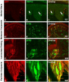
Figure 3. The early Ret+ DRG neurons are the RA mechanoreceptors associated with Meissner corpuscles, Pacinian corpuscles and longitudinal lanceolate endings
A–C: GFP+ fibers do not innervate Merkel cells in the footpad (339/356 {95.2%} of Merkel cells, labeled with the TrpV3 antibody, are not associated with GFP+ axons). The locations of Merkel cells are indicated by white arrows. D–F: GFP+ fibers innervate Meissner corpuscles, visualized by S100 immunostaining, in dermal papillae (114/172 {66.3%} Meissner corpuscles are innervated by GFP+ fibers). G–I: GFP+ fibers form longitudinal lanceolate endings associated with hair follicles, shown by S100 immunostaining (78/184 {42.3%} of hair follicles are associated with GFP+ longitudinal lanceolate endings). J–L: Whole-mount anti-GFP and anti-S100 staining of the periosteum membrane of the fibula. Note that a single GFP+ fiber innervates each Pacinian corpuscle (55/60 {91.7%} of Pacinian corpuscles are innervated by GFP+ fibers). Immunostainings were performed using P14 RetERT2;Tauf(mGFP) mice treated with 4-HT from E10.5 and E12.5. Quantifications were made using four P14 RetERT2;Tauf(mGFP) mice from two litters.
Figure 4
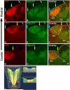
Figure 4. Central projections of early Ret+ DRG neurons innervate layers III through V of the spinal cord
A–D: Central projections of GFP labeled early Ret+ DRG neurons in the upper lumbar level of the spinal cord. Note the presence of GFP+ fibers in both lamina III through V of the spinal cord and the dorsal column (arrow indicates the area which is also outlined by the dotted line). Nociceptor axons innervating spinal cord layers I and II were visualized with CGRP immunostaining (blue) and IB4 binding (green), respectively. E–G: Double staining of GFP and VGLUT1. VGLUT1 labels synapses of the central projections of mechanoreceptors (white dotted line) and proprioceptors (Hughes et al., 2004; Oliveira et al., 2003). H: Dorsal column of the cervical spinal cord. Note that while GFP+ fibers are present in both the gracile (V shape tract, inside the white dotted line) and cuneate fasiculi (area outside the white dotted line), they are greatly enriched in the gracile fasiculus. N=4 from two litters for P14 RetERT2;Tauf(mGFP) mice treated with 4-HT from E10.5 to E12.5.
Figure 5
Figure 5. Modality-specific segregation of mechanosensory axons in dorsal column nuclei
A, D, G: GFP+ fibers innervating the gracile and cuneate nuclei at different rostral to caudal levels of the medulla. The VGLUT1 staining is used to visualize the gracile and cuneate nuclei (B, E, H). The white dotted line outlines the boundary of the gracile nucleus (Gn), and the yellow dotted line outlines the boundary of the cuneate fasiculus and nucleus (Cn) in C, F, I. Note that GFP+ fibers occupy most of the gracile nucleus (I), but are absent from a dorsal segment at the mid-level of the medulla (F). On the other hand, GFP+ fibers do not innervate the caudal-ventral region of the cuneate nucleus (F and I), but project to the dorsal-rostral region of the cuneate nucleus (C). Arrows indicate the boundaries of the gracile and cuneate nuclei. J. Three-dimensional illustration of the pattern of innervation of the gracile and cuneate nuclei by RA mechanoreceptors. This model is based upon the findings reported in Fig. 5A–I and fig S7. The VGLUT1+ zones occupied by GFP+ fibers are showed in yellow. The green zones are unoccupied VGLUT1+ zones. N=3 for 2 month old RetERT2;Tauf(mGFP) mice treated with 4-HT from E10.5 to E12.5.
Figure 6
Figure 6. NRTN-GFRα2/Ret signaling is required for development but not maintenance of Pacinian corpuscles
A–B: Anti-S100 immunostaining of Pacinian corpuscles (white arrows) in the periosteum of the fibula of P14 control and Retf/f;Wnt1Cre mice. Pacinian corpuscles (white arrows) are completely absent (35 ± 5 control vs. 0 mutant, per side) in the Retf/f;Wnt1Cre mice, as visualized using both sections and whole-mount staining (E–F). C–D: Staining of Meissner corpuscles in P14 control and Retf/f;Wnt1Cre mice. Meissner corpuscles (white arrows) appear in normal number in the absence of Ret signaling. However, the morphology of Meissner corpuscles in Retf/f;Wnt1Cre mice is slightly disorganized relative to those found in control mice. E–F: Anti-S100 whole mount immunostaining of Pacinian corpuscles from P14 control and Retf/f;Wnt1Cre mice, Large arrows indicate the interosseous nerve. In the control periosteum membrane, sensory nerves exhibit a tree-like structure on which Pacinian corpuscles are formed (small white arrows indicate a few examples). In contrast, only a few small nerve branches are present (small white arrows), but neither Pacinian corpuscles nor the tree-like nerve branch structures are observed in the Retf/f;Wnt1Cre periosteum membrane, suggesting RA mechanosensory nerves are not present in the absence of Ret. G–H: Staining of longitudinal lanceolate endings (white arrows) in P14 control and Retf/f;Wnt1Cre mice. Longitudinal lanceolate endings are present in Retf/f;Wnt1Cre mice, although they appear morphologically underdeveloped. Higher magnification and quantification of these endings are shown in fig. S11A–C. I–L: H & E staining of Pacinian corpuscles in P14 control (black arrow heads), Retf/f;Wnt1Cre, NRTN null and GFRα2GFP null mice. H & E staining is used here to rule out the potential confounding issue of decreased S100 expression in mutant mice. N≥3 for each mutant genotype.
Figure 7
Figure 7. RA mechanoreceptors require Ret signaling for their assembly into mechanosensory circuits in the spinal cord and medulla
A–B: Synapses of mechanoreceptors, visualized by VGLUT1 staining, in the upper lumbar spinal cord and mid-level medulla of P14 control mice. D–E: Synapses of mechanoreceptors in the upper lumbar spinal cord and mid-level medulla of P14 Retf/f; Wnt1Cre mice. Layers III through V of the spinal cord are outlined by the white dotted line, and Clarke's nucleus is outlined by the yellow dotted line. In the medulla, the gracile nucleus is outlined by the yellow dotted line, and the cuneate nucleus is outlined by the white dotted line. The VGLUT1 staining intensity in layer III through V of spinal cord and gracile nucleus of mutant mice is much lower than that of controls. Quantification of spinal cord staining intensity is shown in panel C (Student's t test, P<0.001 for layers III through V), quantification of the size of the gracile and cuneate nuclei is shown in F (Student's t test, P<0.001 for the gracile nucleus), and quantification of the intensity of the medulla staining is shown in G (Student's t test, P<0.001 for the gracile nucleus). H,L: Anti-GFP staining of RA mechanoreceptor central axonal projections into the spinal cord of P14 RetERT2;Tauf(mGFP) (control) and RetERT/f(CFP) (Ret KO) mice, which were treated with 4-HT at E11.5 and E12.5. Note the reduction in the number of GFP+ axons in RetERT/f(CFP) mice. White arrows indicate the range of projections. Quantification of relative intensity of GFP+ fibers is shown in K. The difference between control and Ret null neurons is statistically significant (Student's t test, P<0.001). I, J: Anti-GFP staining of the dorsal columns of P14 RetERT2;Tauf(mGFP) mice at thoracic (I) and lumbar (J) levels. M,N: Anti-GFP staining of the dorsal columns of P14 RetERT/f(CFP) mice at thoracic (M) and lumbar (N) levels. Many fewer CFP+ fibers are found in the gracile fasciculus of the dorsal column of the mutant (outlined by white dotted lines). Quantification is shown in O. The difference between the control and Ret null neurons is statistically significant (Student's t test, P< 0.001 for the lumbar level, and P<0.01 for the thoracic level). P,Q: Anti-GFP staining of RA mechanosensory fibers innervating the medulla in P14 RetERT2;Tauf(mGFP) mice. T–U: Anti-GFP staining of RA mechanosensory innervation of the medulla in P14 RetERT/f(CFP) mice. The relative area occupied by GFP+ fibers within the gracile (Gn) and cuneat nucleus (Cn) is quantified in S. The difference between the control and Ret null neurons is statistically significant (Student's t test, P<0.01 for the gracile nucleus, and P<0.05 for the cuneate nucleus). R, V: GFP labeled RA mechanosensory DRG neurons in RetERT2;Tauf(mGFP) and RetERT/f(CFP) thoracic DRGs at P14. On average, 8 ± 3 neurons/section are labeled by GFP in RetERT2;Tauf(mGFP) mice while 6 ± 2 neurons/section are labeled by CFP in RetERT/f(CFP) mice. Note that the level of expression of CFP in Ret null neurons is higher than that of control neurons. N=3 for control and Retf/f;Wnt1Cre mice and N=3 for both RetERT2;Tauf(mGFP) and RetERT/f(CFP) mice. 8 to 12 spinal cord sections per animal for each genotype were examined and quantified.
Figure 8
Figure 8. Ret is required autonomously for the formation of the 3rd branch of central RA mechanosensory axons
A–B: DiI labeling of thoracic DRGs of E15.5 RetERT2;Tauf(mGFP) and RetERT/f(CFP) mice. Consistent with published findings (Ozaki and Snider, 1997), mechanoreceptors have already extended central axonal projections to spinal cord layers III through V at E15.5. C: Illustration of RA mechanoreceptor central projections, which can be subdivided into four steps. This illustration is adapted from (Brown, 1981). D: Whole mount anti-GFP staining of a control RetERT2;Tauf(mGFP) DRG from an animal treated with 0.6mg of 4-HT. Note that the 1st order central projections are visible in the DRG. On average, 6 ± 2 GFP+ neurons are labeled per thoracic DRG (12 DRGs in total, N=3 from two separate litters). E: Whole mount anti-GFP staining of a RetERT/f(CFP) DRG from an animal treated with 1mg of 4-HT. Note that 1st order central projections are visible in the DRG. On average, 11 ± 4 CFP+ neurons are labeled per thoracic DRG (20 DRGs in total, N=4 from three separate litters). F: Quantification of labeled DRG neuron number. G: Anti-GFP staining with sagittal thoracic spinal cord sections of RetERT2;Tauf(mGFP) mice. Note that the 3rd order axonal branches (white arrows) originate from rostral-caudal running fibers and penetrate the spinal cord. “*” indicates the position of blood vessels which are autofluorescent and seen in mice of both genotypes. H: Anti-GFP staining of sagittal thoracic spinal cord sections of RetERT/f(CFP) mice. Note that very few 3rd order axonal branches originating from rostral-caudal running fibers are found in these mice. I: Quantification of the numer of 3rd order axonal branches for the two genotypes. On average, 1.48 ± 0.41 3rd order axonal branches are observed in every 200μm rostral-caudal fiber in RetERT2;Tauf(mGFP) control mice (N=3 from two litters, and more than 100 rostralcaudal fibers were measured) while 0.19 ± 0.05 3rd order axonal branches are observed in every 200μm rostral-caudal fiber in RetERT/f(CFP) mutant mice (N=4 from three litters, and more than 100 rostral-caudal fibers were measured). J–K: Anti-GFP staining using horizontal lumbar spinal cord sections taken from RetERT2;Tauf(mGFP) (J) and RetERT/f(CFP) (K) mice. Similar to that observed using sagittal thoracic sections, there are several GFP+ central projections in RetERT2;Tauf(mGFP) control mice at this age, but almost no CFP+ central projections are found in the RetERT/f(CFP) mice.

Similar articles

Cited by

References

    1. Airaksinen MS, Koltzenburg M, Lewin GR, Masu Y, Helbig C, Wolf E, Brem G, Toyka KV, Thoenen H, Meyer M. Specific subtypes of cutaneous mechanoreceptors require neurotrophin-3 following peripheral target innervation. Neuron. 1996;16:287–295. - PubMed
    1. Airaksinen MS, Saarma M. The GDNF family: signalling, biological functions and therapeutic value. Nat Rev Neurosci. 2002;3:383–394. - PubMed
    1. Airaksinen MS, Titievsky A, Saarma M. GDNF family neurotrophic factor signaling: four masters, one servant? Mol Cell Neurosci. 1999;13:313–325. - PubMed
    1. Albrecht F. L. R. a. P. J. The Senses: A Comprehensive Reference. Academic Press; San Diego: 2008. Cutaneous Mechanisms of Tactile Perception: Morphological and Chemical Organization of the Innervation to the Skin; pp. 1–32.
    1. Brown AG. Organization in the spinal cord. Heidelberg; Springer-Verlag; Berlin: New York: 1981.

Publication types

MeSH terms

Substances