. 2009 Dec 24;16(12):1278-89.
doi: 10.1016/j.chembiol.2009.11.015.

Selective inhibitor of proteasome's caspase-like sites sensitizes cells to specific inhibition of chymotrypsin-like sites

Affiliations

Selective inhibitor of proteasome's caspase-like sites sensitizes cells to specific inhibition of chymotrypsin-like sites

Matthew Britton et al. Chem Biol. .

Abstract

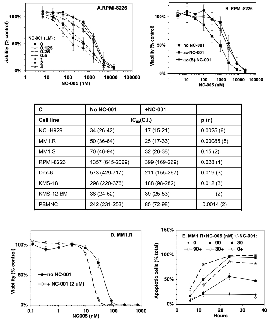
Proteasomes degrade most proteins in mammalian cells and are established targets of anticancer drugs. All eukaryotic proteasomes have three types of active sites: chymotrypsin-like, trypsin-like, and caspase-like. Chymotrypsin-like sites are the most important in protein degradation and are the primary target of most proteasome inhibitors. The biological roles of trypsin-like and caspase-like sites and their potential as cotargets of antineoplastic agents are not well defined. Here we describe the development of site-specific inhibitors and active-site probes of chymotrypsin-like and caspase-like sites. Using these compounds, we show that cytotoxicity of proteasome inhibitors does not correlate with inhibition of chymotrypsin-like sites and that coinhibition of either trypsin-like and/or caspase-like sites is needed to achieve maximal cytotoxicity. Thus, caspase-like and trypsin-like sites must be considered as cotargets of anticancer drugs.


Conflict of interest statement

The authors declare no conflicts of interest.

Figures

Figure 1
Figure 1. Characterization of the novel β5-specific proteasome inhibitor NC-005
A Structures of YU-101 and NC-005. B, C. RPMI-8226 cells were treated with YU-101 and NC-005 for 1h and proteasome activity was measured in cells with ProteasomeGlo assay. Mock-treated cells served as control. Values are averages ± S.E. of two experiments.
Figure 2
Figure 2. Cytotoxicity of the novel proteasome inhibitor NC-005 does not correlate with the inhibition of Chym-L sites
A Overall design of experiments. Cells were treated with inhibitors for 1 h, and then cultured in the absence of the inhibitors for 48 h, when cell viability was assayed with an Alamar Blue. At time points indicated, a fraction of cells was harvested (1, 3, 6, 10, and 23 h) and used for the measurement of apoptosis and of proteasome peptidase activities. B. Recovery of Chym-L activity after 1 h treatment of RPMI-8226 cells with NC-005. Values are averages ± S.E. of two experiments. Recovery of Tr-L and Casp-L activities in RPMI-8226 cells and recovery of all three activities in Dx6 and KMS-18 cells are shown on Fig. S1. C, D. Effect of 1h NC-005 (C) and bortezomib (D) treatment on viability of multiple myeloma cells. Mock-treated cells served as control. Values are averages ± S.E. of 4–6 (C) or 2–3 (D) experiments. E. IC50 (i.e. concentration at which viability was reduced by 50%) was determined by a 4-parameter data fit of dose response curves (C, D) and plotted against specific activity of Chym-L sites. F. Cell viability (from C and D) was plotted against the inhibition of Chym-L (top panel) and Tr-L (middle panel) or Casp-L (bottom panel) sites (values from Table 1). G. Apoptosis of NC-005 treated RPMI-8226 cells. Top panel: caspase-3 activity is expressed in nmole Ac-DEVD-amc cleaved/min/mg protein, and appearance of the cleaved PARP was detected on the Western blot (inset). Bottom panel, percentage of Annexin V-positive apoptotic cells was determined by flow cytometry. Representative results for one out of three different experiments are shown. H. Viability of RPMI-8226, Dox6, and KMS-18 cells (as in C) was plotted against the inhibition of Chym-L (solid lines) and Tr-L (dashed lines) activities at 5 h (top panel) and 10 h (bottom panel) after end of 1 h NC-005-treatment (Fig. S1).
Figure 3
Figure 3. NC-005 derived active site probe and its inactive diastereomer demonstrate that NC-005 is proteasome specific
A RPMI-8226 cells were treated with az-NC-005 active site probe. Activities were measured (values are averages ± S.E. of two experiments) and, in parallel, extracts of these cells were treated with biotinylated phosphane (Fig. 4G) and analyzed by Western blotting. Az-NC-005 reacting polypeptides were visualized by staining with fluorescently labeled streptavidin and identified based on the well-characterized pattern of migration of proteasome’s catalytic subunits (Altun et al., 2005). Endogeneously biotinylated protein of ~70 kDa served as a loading control. B. RPMI-8226 cells were treated with 1.6 µM NC-005 and (S)-NC-005 for 6 h. Proteasome activities were measured immediately after the treatment using peptides substrates (Table); cell viability was measured after subsequent 42 h cultivation in inhibitor-free media.
Figure 4
Figure 4. NC-001 is a highly specific, cell-permeable inhibitor of Casp-L sites
A Structures of NC-001, NC-001- derived probe and it’s inactive diastereomers. B– D. Casp-L (B) and Chym-L and Tr-L activities (C) of the proteasome during continuous treatment of cells with NC-001. Panel D shows all three activities 6 hours after treatment. E. Proteasome peptidase activities in cells treated with az-NC-001 for 6 h. F. Casp-L activity of the proteasome in cells treated for 6 h with az-NC-005 diastereomers. All activity measurements were performed in RPMI-8226 cells and are averages ± S.E. of two independent experiments, in which mock-treated cells served as control. G. Extracts of these cells (i.e., from experiment shown in E and F) were treated with biotinylated phosphane (BioP), separated on 12% gel, transferred on PVDF membrane and probed with fluorescently labeled streptavidin. Polypeptides modified by the probes were identified by incubating the membrane with fluorescently labeled streptavidin. Extracts treated with biotinylated non-site-specific inhibitor Ada-Bio-Ahx3-Leu3-VS were used to identify the migration pattern of proteasome bands (Altun et al., 2005). Endogeneously biotinylated protein of ~70 kDa can be used as a loading control. H. Isolation of az-NC-001-modified subunits by Streptavidin affinity chromatography. Extracts of cells treated overnight with 5 µM az-NC-001 or mock-treated were treated with BioP, passed through a Streptavidin-Sepharose HP column (to remove excess BioP), treated with SDS (to dissociate proteasome subunits), and re-incubated with Sterptavidin-Sepharose. After extensive washing of the beads, specifically bound proteins were eluted with 1 mM biotin in LDS gel loading buffer at 95°C, fractionated on SDS-PAGE, and analyzed by Western blotting with antibodies to proteasome subunits indicated. Percentage of total sample loaded on the gels are indicated. Fig. S2 demonstrates that modification with az-NC-001 and BioP results in the mobility shift of the β1 subunit.
Figure 5
Figure 5. NC-001 sensitizes cells to NC-005
A, B RPMI-8226 cells were treated with NC-005 for 1 h and then split into two equal portions. One was treated with NC-001 (at concentrations indicated, -----A), or az-NC-001, or az-(S)-NC-001 (B) and the other was mock-treated for 48 h, when cell viability was measured with Alamar Blue. Values are averages ± S.E. of two independent experiments. C. NC-001 reduces IC50 of NC-005 (i.e. concentration at which cell viability is reduced by ½ compared to mock-treated cells) in multiple cell lines. IC50 for each individual experiment (performed as in A) were determined by a four-parameter fit of dose-response curves (Figs. S3A (RPMI-8226 cells), 5D (MM1.R cells), and S3B (all other cell lines)). Values in the table are averages (confidence intervals). Results of the t-test are provided in the last column (p-value, n-number of times experiment was repeated). NC-001 was used at 2 µM except in Dox 6 cells where it was at 10 µM. D, E. NC-001 (2 µM) sensitizes MM1.R cells to NC-005 by reducing IC50 (D) and accelerating cell death. Experiment in (E) was performed as in (A), except that at different time points aliquots of cells were collected and analyzed for apoptosis using Annexin V staining (E). Fig. S3A demonstrates that sensitization of cells to NC-005 by NC-001 does not depend on the order of treatment with these reagents. Fig. S3B demonstrates sensitization of other myeloma cells lines to NC-005 by NC-001.
Figure 6
Figure 6. Effect of NC-001 on recovery of proteasome activity in NC-005-treated cells
A. MM1.R cells were treated with NC-001 in the presence or absence of 2 µM NC-001 for 1h, followed by incubation in media in the presence or absence of NC-001 respectively. At times indicated, some cells were harvested and proteasome activity was measured in extracts with fluorogenic peptide substrates and normalized per mg total protein (determined by Bradford). No inhibition of Tr-L activity was observed. B. RPMI-8226 cells were treated with NC-005 for 1h, followed by the NC-001 or mock-treatment. At times indicated proteasome activity was measured with ProteasomeGlo assay and normalized per number cell at time zero. Time-dependent decrease in Tr-L and Casp-L activity in RPMI-8226 cells treated with higher concentrations of NC-005 reflects time-dependent decrease in the number of cells due to inhibition of cell proliferation or cell death. Solid lines, no NC-001; dashed lines, with NC-001. Values are averages ± S.E. of two independent experiments. Fig. S4A demonstrates effects of NC-001 on recovery of proteasome activity in NC-005 treated Dox6 and KMS-18 cells. Fig. S4B demonstrates that ProteasomeGlo and fluorogenic substrate assays give identical results.

Similar articles

Cited by

References

    1. Adams J. The proteasome: a suitable antineoplastic target. Nat Rev Cancer. 2004;4:349–360. - PubMed
    1. Adams J, Palombella VJ, Sausville EA, Johnson J, Destree A, Lazarus DD, Maas J, Pien CS, Prakash S, Elliott PJ. Proteasome inhibitors: a novel class of potent and effective antitumor agents. Cancer Res. 1999;59:2615–2622. - PubMed
    1. Altun M, Galardy PJ, Shringarpure R, Hideshima T, LeBlanc R, Anderson KC, Ploegh HL, Kessler BM. Effects of PS-341 on the activity and composition of proteasomes in multiple myeloma cells. Cancer Res. 2005;65:7896–7901. - PubMed
    1. Arendt CS, Hochstrasser M. Identification of the yeast 20S proteasome catalytic centers and subunit interactions required for active-site formation. Proc Natl Acad Sci. 1997;94:7156–7161. - PMC - PubMed
    1. Berkers CR, Verdoes M, Lichtman E, Fiebiger E, Kessler BM, Anderson KC, Ploegh HL, Ovaa H, Galardy PJ. Activity probe for in vivo profiling of the specificity of proteasome inhibitor bortezomib. Nat Methods. 2005;2:357–362. - PubMed

Publication types

LinkOut - more resources