. 2010 May;171(1):8-16.
doi: 10.1016/j.molbiopara.2009.12.006. Epub 2010 Jan 11.

Levamisole and ryanodine receptors. II: An electrophysiological study in Ascaris suum

Affiliations

Levamisole and ryanodine receptors. II: An electrophysiological study in Ascaris suum

Sreekanth Puttachary et al. Mol Biochem Parasitol. 2010 May.

Abstract

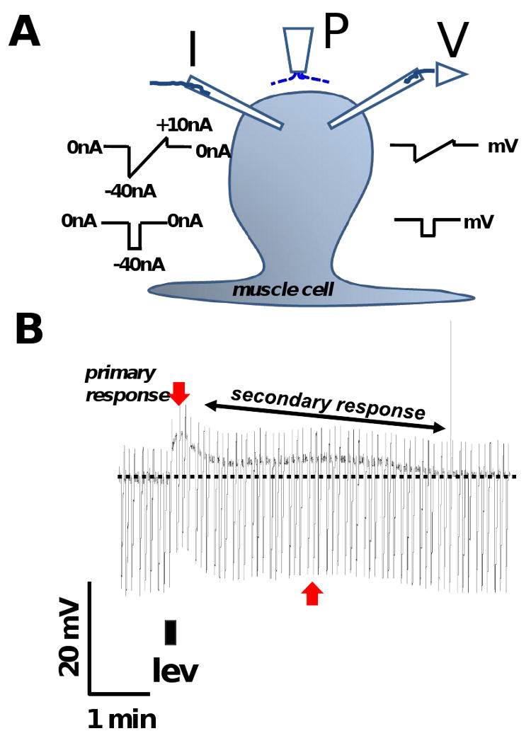
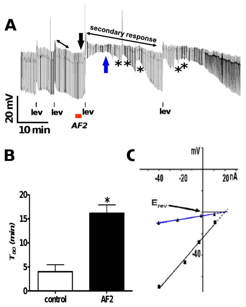
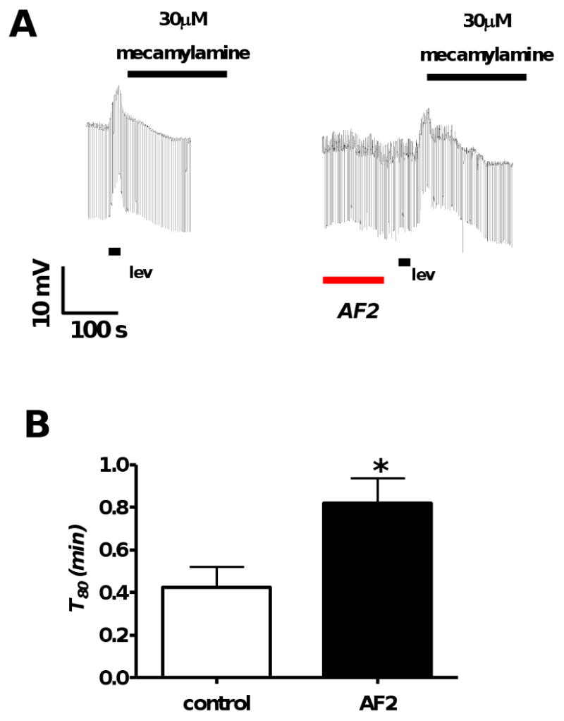
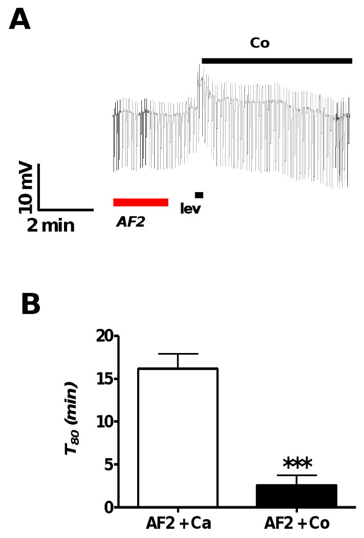
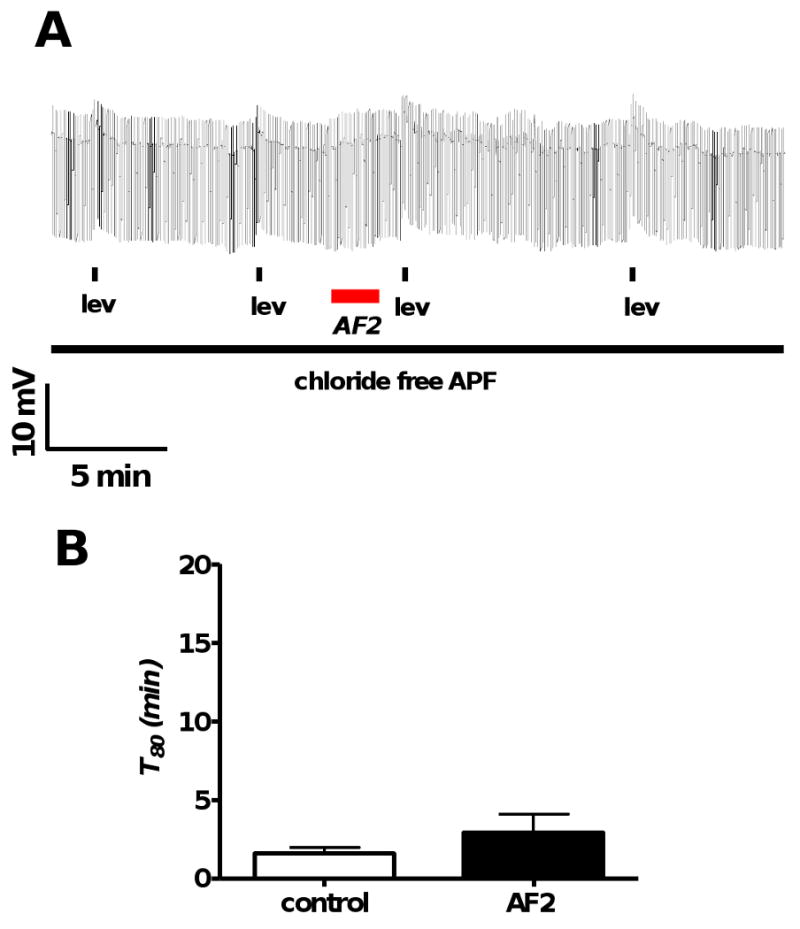
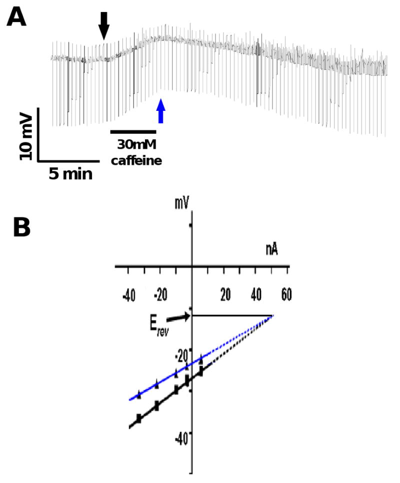
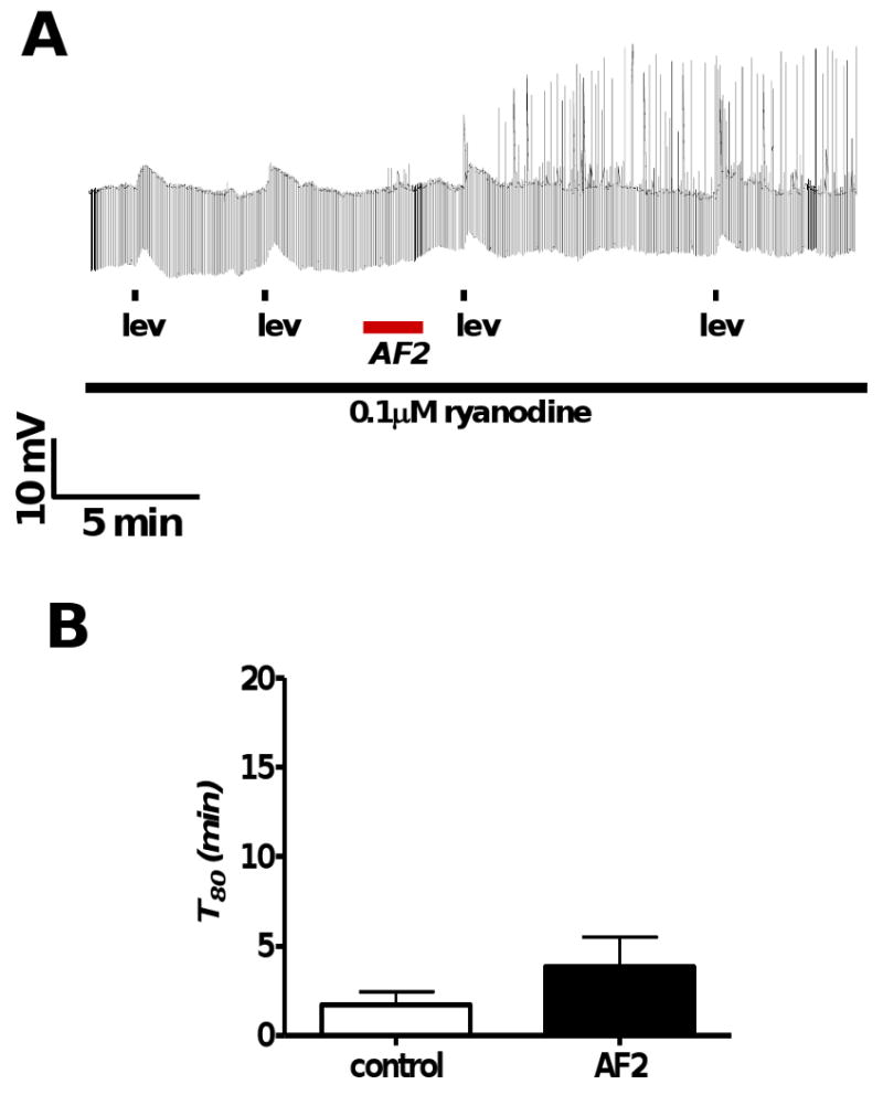
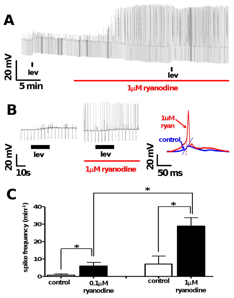
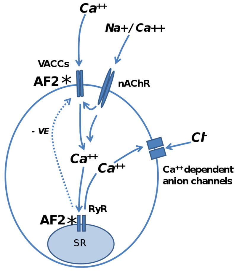
Resistance to antinematodal drugs like levamisole has increased and there is a need to understand what factors affect the responses to these anthelmintics. In our previous study, we examined the role of ryanodine receptors in muscle contraction pathways. Here we have examined interactions of levamisole receptors, ryanodine receptors (RyRs), the excitatory neuropeptide AF2, and coupling to electrophysiological responses. We examined the effects of a brief application of levamisole on Ascaris suum body muscle under current-clamp. The levamisole responses were characterized as an initial primary depolarization, followed by a slow secondary depolarizing response. We examined the effects of AF2 (KHEYLRFamide), 1 microM applied for 2 min. We found that AF2 potentiated the secondary response to levamisole and had no significant effect on the primary depolarization. Further, the reversal potentials observed during the secondary response suggested that more than one ion was involved in producing this potential. AF2 potentiated the secondary response in the presence of 30 microM mecamylamine suggesting the effect was independent of levamisole sensitive acetylcholine receptors. The secondary response, potentiated by AF2, appeared to be dependent on cytoplasmic events triggered by the primary depolarization. Ion-substitution experiments showed that the AF2 potentiated secondary response was dependent on extracellular calcium and chloride suggesting a role for the calcium-activated anion channel. Caffeine mimicked the AF2 potentiated secondary response and 0.1 microM ryanodine inhibited it. 1.0 microM ryanodine increased spiking showing that it affected membrane excitability. A model is proposed showing ryanodine receptors mediating effects of AF2 on levamisole responses.


Figures

Fig. 1
Fig. 1
A: Diagram showing the placement of the two micropipettes used for current-clamp and position of the micro perfusion system for continuous perfusion and application of drugs. P: microperfusion pipette. I: current-injecting electrode, injects ramp currents or step currents. V: voltage-recording electrode. B: Representative trace showing the levamisole response and its 2 components in APF-Ringer namely, a primary depolarization and a secondary depolarizing response (secondary response). The darkest line of the recording is the membrane potential and the downward transients are the responses to injected current. The rapid primary depolarization (downward red arrow) is followed by a slow secondary response (red vertical arrow and oblique black double arrow). 1 μM levamisole was applied for 10 s as indicated by the filled rectangle below the trace. The discontinuous horizontal line indicates the original position of the resting membrane potential. The width of the trace is a reflection of membrane conductance; it gets narrower as membrane-ion channels open. The duration of the secondary response (T80) was measured as the time taken (min) for the peak primary depolarization to decline by 80%.
Fig. 2
Fig. 2
A: Representative current-clamp trace showing AF2 potentiating the secondary response to levamisole. There are two applications of levamisole (1 μM) before and after AF2 treatment. The control levamisole applications are followed by a 2 min application of AF2 (1 μM) with a brief wash (1 min), subsequently; there are two test levamisole applications. The double headed black arrows represent the secondary response before and AF2 treatment. * represents waves of conductance change during the secondary response. B: Bar graph comparing T80 (min) before and after AF2 treatment in APF-Ringer. The secondary response to levamisole application was significantly increased after AF2 treatment as indicated by the increase in T80 (Fig 2B, n = 4, p < 0.05, paired t-test). C: The black line and downward arrow show the control current-voltage plot before the test levamisole application and the blue upward arrow and line show the current-voltage plot during the at the secondary peak from Fig. 2A. Plots were fitted by linear regression. The reversal potential, estimated by extrapolating the two current-voltage plots are shown. The reversal potential, Erev, was -20 mV.
Fig. 3
Fig. 3
A: Representative current-clamp traces where mecamylamine (30 μM), a nAChR antagonist, was applied immediately after the end of levamisole application in the control (n = 4) and the test (n = 5) recordings. The first trace shows a control and the second trace shows the test response after AF2. B: Bar graph comparing T80 controls and AF2 test pre-treatments in the presence of mecamylamine. AF2 potentiated the duration of levamisole secondary response T80, in the presence of mecamylamine (p < 0.05, unpaired t-test).
Fig. 4
Fig. 4
A: Representative current-clamp trace showing the lack of an AF2 potentiated secondary response following replacement of calcium with cobalt APF-Ringer following the end of levamisole application (1 μM). B: Bar graph comparing mean durations of secondary depolarizations, T80, from different preparations recorded after AF2 treatment in the presence and absence of calcium (calcium replaced using cobalt APF-Ringer). Calcium substitution caused a significant reduction in the duration of the secondary response (p < 0.001, n = 4, unpaired t-test).
Fig. 5
Fig. 5
A: Representative current-clamp trace showing levamisole (1 μM) applications before and after AF2 treatment in the chloride free in APF-Ringer. B: Bar graph comparing T80 before and after AF2 treatment. In the absence of extracellular chloride, T80 in control and test responses were not significantly different (p > 0.05, n = 5, paired t-test). AF2 did not potentiate the levamisole secondary response in the absence of chloride as indicated by T80 measurements.
Fig. 6
Fig. 6
A: Current-clamp trace showing the effect of caffeine (30 mM) on the membrane potential and conductance. Note that the application of caffeine produced a slow depolarization associated with an increase in conductance. B: The membrane potential responses to the injected ramp currents fitted with linear regression before application of caffeine (black arrow Fig. 6 A) and at the peak depolarization (blue arrow Fig 6A) in the IV plot. The reversal potential, Erev, estimated after extrapolating the membrane potential responses was -12 mV.
Fig. 7
Fig. 7
A: Representative current-clamp trace showing levamisole (1 μM) applications before and after AF2 treatment in the presence of 0.1 μM ryanodine. B: Bar graph depicting T80 (min) in control and test applications. In the presence of 0.1 μM ryanodine, T80 in control and test responses were not significantly different (p > 0.05, n = 4, paired t-test). AF2 did not significantly potentiate the levamisole secondary response in the presence of ryanodine, as indicated by T80 measurements.
Fig. 8
Fig. 8
A: Representative current-clamp trace showing levamisole (1 μM) application before and during ryanodine treatment (1 μM). B: Representative trace of spikes seen at higher time resolution during levamisole application before and in the presence of 1 μM ryanodine. The recordings show an increase the spike amplitudes (left) and an increase in the gradient of the rising phase of the spikes (blue increased in red: right) in the presence of ryanodine. C: Bar graphs showing the mean ± S.E. spike frequency during the depolarizing phase of the response to levamisole before and in the presence of 0.1 μM ryanodine and in separate experiments 1 μM ryanodine. Treatment with 0.1 μM ryanodine significantly increased spiking from 0.8 ± 0.8 min-1 to 6 ± 2.1 min-1 (n = 4, p < 0.05, paired t-test). Treatment with 1 μM ryanodine significantly increased spiking from 7.2 ± 4.6 min-1 to 28.9 ± 4.8 min-1 (n = 6, p < 0.05, paired t-test). Spiking frequencies at 1 μM were also significantly greater than at 0.1 μM (p<0.01, unpaired t-test) demonstrating that the effect of ryanodine on spiking was concentration dependent.
Fig. 9
Fig. 9
Proposed model and sites of action whereby AF2 modulates the responses to levamisole [1]. The primary depolarization follows levamisole binding to nAChRs and their opening to allow Ca++ and Na+ to enter the cell. The levamisole secondary response is initiated by the primary depolarization and involves activation of voltage-gated calcium channels (VACCs), ryanodine receptors (RyRs) and calcium-activated anion channels. VACCs are activated by the primary depolarization and allow more calcium to enter the cell. Increased intracellular calcium triggers calcium induced calcium release (CICR) from the sarcoplasmic reticulum (SR) and are gated by the ryanodine channels (RyRs). The calcium-activated anion channels are also activated during the cytoplasmic rise in calcium concentration. The calcium induced calcium release can inhibit the voltage-activated calcium channels (VACCs) as a negative feedback (- ve). *: AF2 potentiates the levamisole secondary responses by increasing voltage-activated calcium entry through VACCs [19] and; * by sensitizing the RyRs to release more calcium from the sarcoplasmic reticulum in response to the calcium entry through the nAChRs and VACCs.

Similar articles

Cited by

References

    1. Trailovic SM, et al. Brief application of AF2 produces long lasting potentiation of nAChR responses in Ascaris suum. Mol Biochem Parasitol. 2005;139:51–64. - PubMed
    1. Albonico M, Engels D, Savioli L. Monitoring drug efficacy and early detection of drug resistance in human soil-transmitted nematodes: a pressing public health agenda for helminth control. Int J Parasitol. 2004;34:1205–10. - PubMed
    1. Albonico M, et al. Development of the egg hatch assay for detection of anthelmintic resistance in human hookworms. Int J Parasitol. 2005;35:803–11. - PubMed
    1. Jackson F. Anthelmintic resistance-the state of play. Br Vet J. 1993;149:123–38. - PubMed
    1. Kaplan RM. Drug resistance in nematodes of veterinary importance: a status report. Trends Parasitol. 2004;20:477–81. - PubMed

Publication types

MeSH terms

LinkOut - more resources