. 2010 Mar;119(1-2):73-83.
doi: 10.1016/j.jsbmb.2010.01.001. Epub 2010 Jan 11.

1,25(OH)2vitamin D3 inhibits cell proliferation by promoting cell cycle arrest without inducing apoptosis and modifies cell morphology of mesenchymal multipotent cells

Affiliations

1,25(OH)2vitamin D3 inhibits cell proliferation by promoting cell cycle arrest without inducing apoptosis and modifies cell morphology of mesenchymal multipotent cells

Jorge N Artaza et al. J Steroid Biochem Mol Biol. 2010 Mar.

Abstract

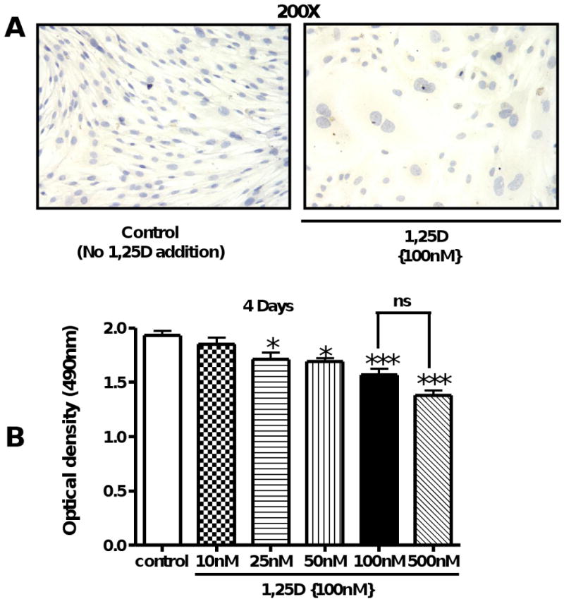
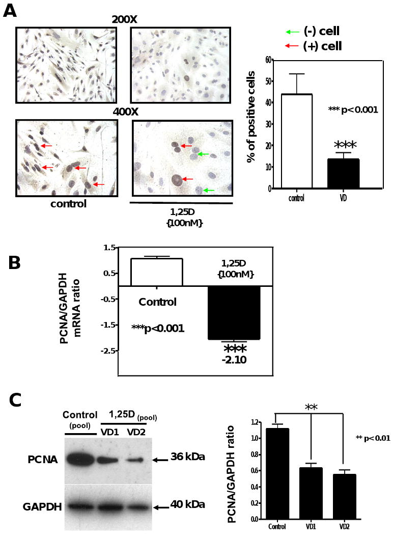
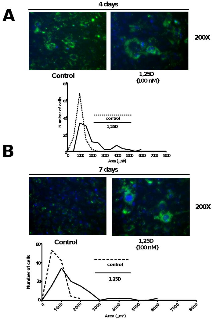
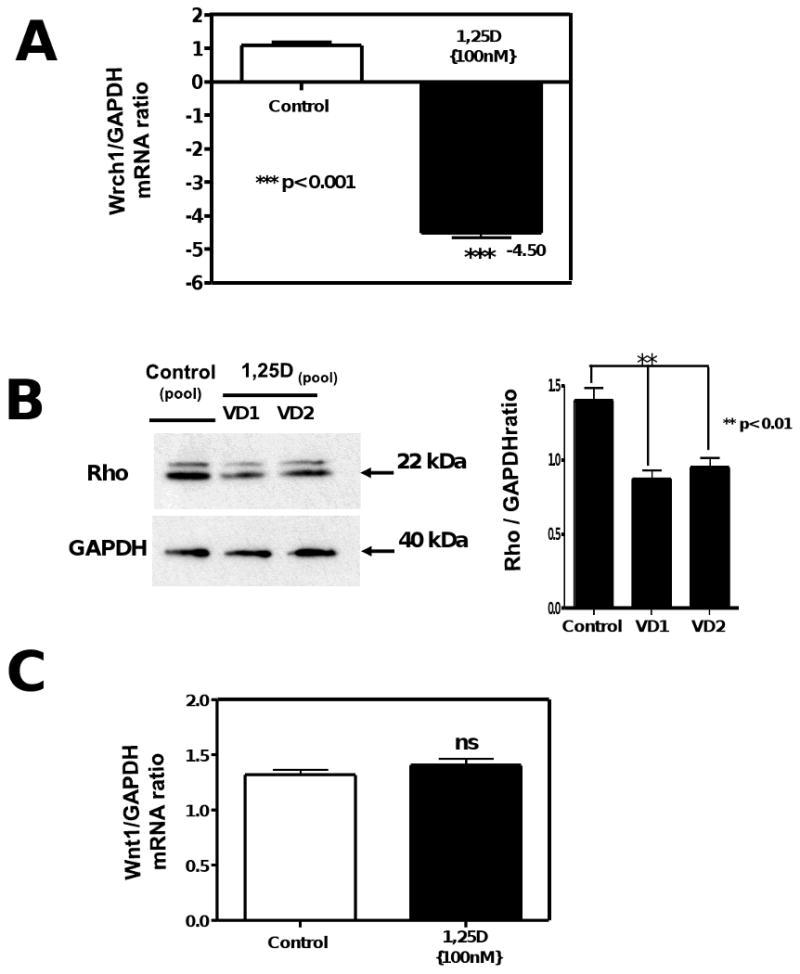
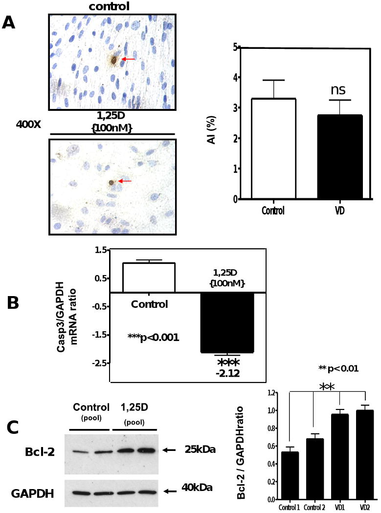
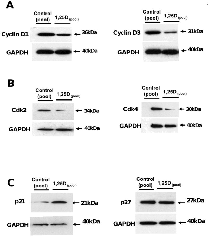
The vitamin D receptor (VDR) and its ligand 1,25D play an important role in regulating cell growth and cell fate. We examined the effect of 1,25D on cell morphology, cell proliferation, cell cycle progression and apoptosis on mesenchymal multipotent cells. Multipotent cells were treated with and without 1,25D in a time- and dose-dependent manner. Changes in cell morphology were evaluated by a green fluorescence fluorocrome. Cell proliferation was determined by the Formazan assay and PCNA antigen expression. The expression of genes related to the cell cycle was analyzed by DNA microarrays, RT(2)PCR arrays and western blots. Apoptosis was evaluated by TUNEL assay, and the expression of pro- and anti-apoptotic related genes by RT(2)PCR arrays and western blots. 1,25D inhibited cell proliferation, induced cell cycle arrest, and promoted accumulation of cells in G0/G1 phase without inducing apoptosis. An increase in cell size was associated with a decrease in the GTPase Rho and the atypical Rho family GTPase Rhou/Wrch-1 expression without inducing Wnt-1 expression. Survivin expression was also increased and may represent a novel 1,25D-mediated pathway regulating tissue injury and fibrosis. The data provide a mechanistic explanation for the anti-proliferative and anti-apoptotic properties of 1,25D in mesenchymal multipotent cells.


Figures

Figure 1
Figure 1. 1,25D inhibits cell proliferation in Multipotent mesenchymal C3H 10T1/2 in a dose dependent manner
(A) Cells treated with or without 1,25D (100 nM) for 4 days were fixed with 2% p-formaldehyde and counterstained with hematoxyline (200×). (B) C3H 10T1/2 cells primed with 5′-azacytidine (20 μM) at an initial number of 4,000 cells/well were incubated for 4 days, as follows: control: no addition of 1,25D and 1,25D: increasing concentrations of 1,25D from 10 – 500 nM. Results are expressed as Mean±SEM. *P<0.05; ***P<0.001
Figure 2
Figure 2. 1,25D downregulates the expression of proliferating cell nuclear antigen (PCNA)
Cells treated as in Figure 1 were incubated with or without 100nM of 1,25D for 4 days. Immunocytochemistry for PCNA were performed at the end of the incubation time (A). Mean±SEM corresponds to experiments done in triplicates of the percentage of positive cells (brown nuclear staining) by quantitative image analysis. ***p<0.001. (B) Total RNA isolation followed by real time PCR was applied in other aliquots for the 4 days incubations normalized by GAPDH housekeeping gene. Mean±SEM corresponds to experiments done in triplicates ***p<0.001. (C) Western blots analysis was performed for extracts from the 4 days incubation time (left) and the corresponding densitometry analysis (right). 200× and 400×.
Figure 3
Figure 3. 1,25D decreases cell number and increases cell size of multipotent mesenchymal cells
Cells treated as in Figure 1 were preincubated with Green Fluorescent Cell Linker (PKH2) and counterstaing with DAPI. Next day, cells were incubating with or without 1,25D for 4 and 7 days respectively. Representative pictures are presented for control (No 1,25D addition) and 1,25D (100nM) at 4 days (A upper panel) and at 7 days (B, upper panel) with the corresponding histograms showing the cell size distribution and expressing the number of cells per area (μm2). (A and B, lower panels)
Figure 4
Figure 4. 1,25D regulates cell size through downregulation of the GTPase Rho and the atypical rho family GTPase Rhou/Wrcth-1 without intervention of Wnt1
Total RNA isolation from cells treated as in Figure 2, was subjected to real time PCR for Wrcth1 normalized by GAPDH housekeeping gene (A). Mean±SEM corresponds to experiments done in triplicates ***p<0.001. (B) Western blots analysis was performed for extracts from the 4 days incubation time for Rho normalized with GAPDH with the correspondent densitometric analysis. In another set of aliquots the RNA samples were utilized for real time PCR for Wnt1 normalized by GAPDH housekeeping gene (C).
Figure 5
Figure 5. 1,25D exerts an antiapoptotic effect in C3H 10T1/2 multipotent mesenchymal cells
Cells treated as in Figure 2, were seeded in 8-well chamber slides for TUNEL assay or in 6-well plates for RNA and protein isolation. At the end of the incubation period, samples were subjected to TUNEL assay where the apoptotic index (AI) was obtained for control and VD incubated samples (A). In another set of samples, total RNA isolation was followed by real time PCR for Caspase 3 normalized by GAPDH housekeeping gene. Mean±SEM corresponds to experiments done in triplicates ***p<0.001. (B) Western blots analysis was performed for extracts from the 4 days incubation time for Bcl-2 normalized with GAPDH with the corresponding densitometric analysis (C).
Figure 6
Figure 6. 1,25D promotes cell cycle arrest in C3H 10T1/2 mesenchymal multipotent cells by decreasing the expression of cyclins, cyclin dependent kinases (Cdk) and Cdki (cyclin dependent kinases inhibitors)
Cells treated as in Figure 2, were seeded on 6-well plates for protein isolation Western blots analysis was performed for extracts from the 4 days incubation time for Cyclin D1 and D3 (A), for Cdk2 and Cdk4 (B) and p21 and p27 (C) all of them normalized for housekeeping genes with GAPDH.

References

    1. Feldman D, Pike JW, Glorieux FH. Vitamin D. 2nd. Elsevier Academic Press; 2005. Chapter 11.
    1. Dusso AS, Brown AJ. Mechanism of vitamin D action and its regulation. Am J Kidney Dis. 1998;32:S13–S24. - PubMed
    1. Dusso AS, Brown AJ, Slatopolsky E. Vitamin D. Am J Physiol Renal Physiol. 2005;289:F8–F28. - PubMed
    1. Zehnder D, Bland R, Williams MC, McNinch RW, Howie AJ, Stewart PM, Hewison M. Extrarenal expression of 25-hydroxyvitamin d(3)-1 alpha-hydroxylase. J Clin Endocrinol Metab. 2001;86(2):888–94. - PubMed
    1. Carlberg C, Quack M, Herdick M, Bury Y, Polly P, Toell A. Central role of VDR conformations for understanding selective actions of vitamin D(3) analogues. Steroids. 2001;66:213–221. - PubMed

Publication types

MeSH terms

LinkOut - more resources