

The KATP channel is a molecular sensor of atrophy in skeletal muscle
- PMID: 20064856
- PMCID: PMC2834937
- DOI: 10.1113/jphysiol.2009.185835
The KATP channel is a molecular sensor of atrophy in skeletal muscle
Abstract
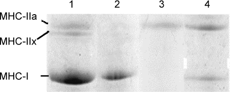
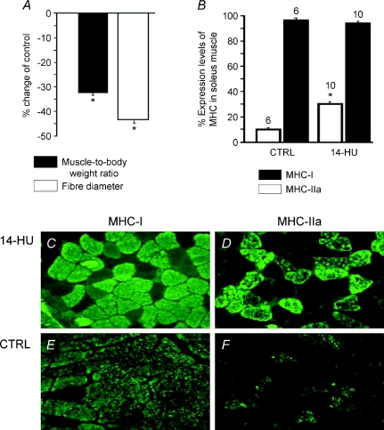
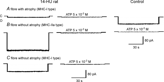
The involvement of ATP-sensitive K(+) (K(ATP)) channels in the atrophy of slow-twitch (MHC-I) soleus (SOL) and fast-twitch (MHC-IIa) flexor digitorum brevis (FDB) muscles was investigated in vivo in 14-day-hindlimb-unloaded (14-HU) rats, an animal model of disuse, and in vitro in drug-induced muscle atrophy. Patch-clamp and gene expression experiments were performed in combination with measurements of fibre diameters used as an index of atrophy, and with MHC labelling in 14-HU rats and controls. A down-regulation of K(ATP) channel subunits Kir6.2, SUR1 and SUR2B with marked atrophy and incomplete phenotype transition were observed in SOL of 14-HU rats. The observed changes in K(ATP) currents were well correlated with changes in fibre diameters and SUR1 expression, as well as with MHC-IIa expression. Half of the SOL fibres of 14-HU rats had reduced diameter and K(ATP) currents and were labelled by MHC-I antibodies. Non-atrophic fibres were labelled by MHC-IIa (22%) antibodies and had enhanced K(ATP) currents, or were labelled by MHC-I (28%) antibodies but had normal current. FDB was not affected in 14-HU rats and this is related to the high expression/activity of Kir6.2/SUR1 subunits characterizing this muscle phenotype. The long-term incubation of the control muscles in vitro with the K(ATP) channel blocker glibenclamide (10(6)m) reduced the K(ATP) currents with atrophy and these effects were prevented by the K(ATP) channel opener diazoxide (10(4)m). The in vivo down-regulation of SUR1, and possibly of Kir6.2 and SUR2B, or their in vitro pharmacological blockade activates atrophic signalling in skeletal muscle. All these findings suggest a new role for the K(ATP) channel as a molecular sensor of atrophy.
Figures
Similar articles
-
Dual response of the KATP channels to staurosporine: a novel role of SUR2B, SUR1 and Kir6.2 subunits in the regulation of the atrophy in different skeletal muscle phenotypes.Biochem Pharmacol. 2014 Sep 15;91(2):266-75. doi: 10.1016/j.bcp.2014.06.023. Epub 2014 Jul 3. Biochem Pharmacol. 2014. PMID: 24998494
-
Effects of ZD0947, a novel and potent ATP-sensitive K+ channel opener, on smooth muscle-type ATP-sensitive K+ channels.Eur J Pharmacol. 2016 Nov 15;791:773-779. doi: 10.1016/j.ejphar.2016.09.038. Epub 2016 Sep 29. Eur J Pharmacol. 2016. PMID: 27693800
-
Reduced expression of Kir6.2/SUR2A subunits explains KATP deficiency in K+-depleted rats.Neuromuscul Disord. 2008 Jan;18(1):74-80. doi: 10.1016/j.nmd.2007.07.009. Epub 2007 Sep 6. Neuromuscul Disord. 2008. PMID: 17825556
-
Targeting hypertension with a new adenosine triphosphate-sensitive potassium channel opener iptakalim.J Cardiovasc Pharmacol. 2010 Sep;56(3):215-28. doi: 10.1097/FJC.0b013e3181e23e2b. J Cardiovasc Pharmacol. 2010. PMID: 20410832 Review.
-
SUR1-TRPM4 channels, not KATP, mediate brain swelling following cerebral ischemia.Neurosci Lett. 2020 Jan 23;718:134729. doi: 10.1016/j.neulet.2019.134729. Epub 2019 Dec 31. Neurosci Lett. 2020. PMID: 31899311 Free PMC article. Review.
Cited by
-
Nerve Growth Factor, Brain-Derived Neurotrophic Factor and Osteocalcin Gene Relationship in Energy Regulation, Bone Homeostasis and Reproductive Organs Analyzed by mRNA Quantitative Evaluation and Linear Correlation Analysis.Front Physiol. 2016 Oct 13;7:456. doi: 10.3389/fphys.2016.00456. eCollection 2016. Front Physiol. 2016. PMID: 27790153 Free PMC article.
-
Database search of spontaneous reports and pharmacological investigations on the sulfonylureas and glinides-induced atrophy in skeletal muscle.Pharmacol Res Perspect. 2014 Feb;2(1):e00028. doi: 10.1002/prp2.28. Epub 2014 Mar 3. Pharmacol Res Perspect. 2014. PMID: 25505577 Free PMC article.
-
Commentary: A BK (Slo1) channel journey from molecule to physiology.Front Pharmacol. 2017 Apr 5;8:188. doi: 10.3389/fphar.2017.00188. eCollection 2017. Front Pharmacol. 2017. PMID: 28424624 Free PMC article. No abstract available.
-
The Impact of Glucose-Lowering Drugs on Sarcopenia in Type 2 Diabetes: Current Evidence and Underlying Mechanisms.Cells. 2021 Aug 1;10(8):1958. doi: 10.3390/cells10081958. Cells. 2021. PMID: 34440727 Free PMC article. Review.
-
The Impact of Antidiabetic Agents on Sarcopenia in Type 2 Diabetes: A Literature Review.J Diabetes Res. 2020 Jul 9;2020:9368583. doi: 10.1155/2020/9368583. eCollection 2020. J Diabetes Res. 2020. PMID: 32695832 Free PMC article. Review.
References
-
- Andersen JL, Aagaard P. Myosin heavy chain IIX overshoot in human skeletal muscle. Muscle Nerve. 2000;23:1095–1104. - PubMed
-
- Debska G, Kicinska A, Skalska J, Szewczyk A, May R, Elger CE, Kunz WS. Opening of potassium channels modulates mitochondrial function in rat skeletal muscle. Biochim Biophys Acta. 2002;1556:97–105. - PubMed
-
- Desaphy JF, Pierno S, Léoty C, George AL, Jr, De Luca A, Conte Camerino D. Skeletal muscle disuse induces fibre type-dependent enhancement of Na+ channel expression. Brain. 2001;124:1100–1113. - PubMed
-
- Desaphy J-F, Pierno S, Liantonio A, De Luca A, Didonna MP, Frigeri A, Nicchia GP, Svelto M, Camerino D, Zallone A, Conte Camerino D. Recovery of the soleus muscle after short- and long-term disuse induced by hindlimb unloading: effects on the electrical properties and myosin heavy chain profile. Neurobiol Dis. 2005;18:356–365. - PubMed
Publication types
MeSH terms
Substances
LinkOut - more resources
Full Text Sources
Research Materials