Contribution of afferent feedback and descending drive to human hopping
- PMID: 20064857
- PMCID: PMC2834939
- DOI: 10.1113/jphysiol.2009.182709
Contribution of afferent feedback and descending drive to human hopping
Abstract
During hopping an early burst can be observed in the EMG from the soleus muscle starting about 45 ms after touch-down. It may be speculated that this early EMG burst is a stretch reflex response superimposed on activity from a supra-spinal origin. We hypothesised that if a stretch reflex indeed contributes to the early EMG burst, then advancing or delaying the touch-down without the subject's knowledge should similarly advance or delay the burst. This was indeed the case when touch-down was advanced or delayed by shifting the height of a programmable platform up or down between two hops and this resulted in a correspondent shift of the early EMG burst. Our second hypothesis was that the motor cortex contributes to the first EMG burst during hopping. If so, inhibition of the motor cortex would reduce the magnitude of the burst. By applying a low-intensity magnetic stimulus it was possible to inhibit the motor cortex and this resulted in a suppression of the early EMG burst. These results suggest that sensory feedback and descending drive from the motor cortex are integrated to drive the motor neuron pool during the early EMG burst in hopping. Thus, simple reflexes work in concert with higher order structures to produce this repetitive movement.
Figures

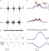
 and the end indicates peak of the early EMG burst and thus the length of the bar indicates the duration between those events. It can be seen that the time between
  and the peak of the early EMG burst remains the same in the three conditions. Only the time of
  is displayed in the stimulated condition of the TMS protocol since the position of the peak is concealed by the after-effects of the suppression. In C‘TMS stimulus’ indicates the time point where the TMS stimulus is delivered. ‘Suppression’ represents the average timing and duration of the suppression. By comparing it with the control condition in Fig. 3B), it can be seen that the suppression is timed just prior to the peak of the early EMG burst.
 . B, ensemble average of 25 sweeps for the conditions ‘Up’, ‘Level’ and ‘Down’. All averaged trials are aligned to the crossing of the light barrier. Note that the platform in the ‘Up’ position causes a shift in the signals for ankle angle, ground reaction force and the EMG burst in SOL ahead in time whereas the ‘Down’ position delays these signals in time.
 or EMG peak) is delayed compared to the level position (platform down); negative numbers are situations in which those events are shifted ahead (platform up).
Similar articles
-
Age-related differences in the neural regulation of stretch-shortening cycle activities in male youths during maximal and sub-maximal hopping.J Electromyogr Kinesiol. 2012 Feb;22(1):37-43. doi: 10.1016/j.jelekin.2011.09.008. Epub 2011 Oct 14. J Electromyogr Kinesiol. 2012. PMID: 22000942
-
Contribution of afferent feedback to the soleus muscle activity during human locomotion.J Neurophysiol. 2005 Jan;93(1):167-77. doi: 10.1152/jn.00283.2004. Epub 2004 Sep 8. J Neurophysiol. 2005. PMID: 15356177
-
Reflex facilitation during the stretch-shortening cycle.J Electromyogr Kinesiol. 2000 Jun;10(3):179-87. doi: 10.1016/s1050-6411(00)00007-9. J Electromyogr Kinesiol. 2000. PMID: 10818339
-
Contributions to the understanding of gait control.Dan Med J. 2014 Apr;61(4):B4823. Dan Med J. 2014. PMID: 24814597 Review.
-
Cutaneous silent periods.Muscle Nerve. 2003 Oct;28(4):391-401. doi: 10.1002/mus.10447. Muscle Nerve. 2003. PMID: 14506711 Review.
Cited by
-
The Anticipation of Gravity in Human Ballistic Movement.Front Physiol. 2021 Mar 17;12:614060. doi: 10.3389/fphys.2021.614060. eCollection 2021. Front Physiol. 2021. PMID: 33815134 Free PMC article.
-
Fractionation of the visuomotor feedback response to directions of movement and perturbation.J Neurophysiol. 2014 Nov 1;112(9):2218-33. doi: 10.1152/jn.00377.2013. Epub 2014 Aug 6. J Neurophysiol. 2014. PMID: 25098965 Free PMC article.
-
Triceps surae short latency stretch reflexes contribute to ankle stiffness regulation during human running.PLoS One. 2011;6(8):e23917. doi: 10.1371/journal.pone.0023917. Epub 2011 Aug 22. PLoS One. 2011. PMID: 21887345 Free PMC article.
-
Processing information related to centrally initiated locomotor and voluntary movements by feline spinocerebellar neurones.J Physiol. 2011 Dec 1;589(Pt 23):5709-25. doi: 10.1113/jphysiol.2011.213678. Epub 2011 Sep 19. J Physiol. 2011. PMID: 21930605 Free PMC article.
-
Effects of conditioning hops on drop jump and sprint performance: a randomized crossover pilot study in elite athletes.BMC Sports Sci Med Rehabil. 2016 Jan 30;8:1. doi: 10.1186/s13102-016-0027-z. eCollection 2016. BMC Sports Sci Med Rehabil. 2016. PMID: 26835128 Free PMC article.
References
-
- Agarwal GC, Gottlieb GL. Effect of vibration of the ankle stretch reflex in man. Electroencephalogr Clin Neurophysiol. 1980;49:81–92. - PubMed
Publication types
MeSH terms
LinkOut - more resources
Full Text Sources

