. 2010 Mar 19;285(12):8719-32.
doi: 10.1074/jbc.M109.077081. Epub 2010 Jan 11.

Epithelial cell adhesion molecule regulation is associated with the maintenance of the undifferentiated phenotype of human embryonic stem cells

Affiliations

Epithelial cell adhesion molecule regulation is associated with the maintenance of the undifferentiated phenotype of human embryonic stem cells

Tung-Ying Lu et al. J Biol Chem. .

Abstract

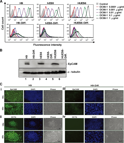
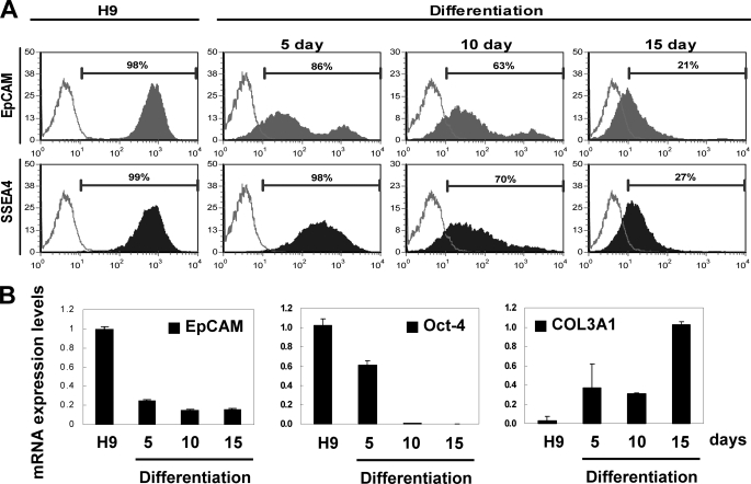
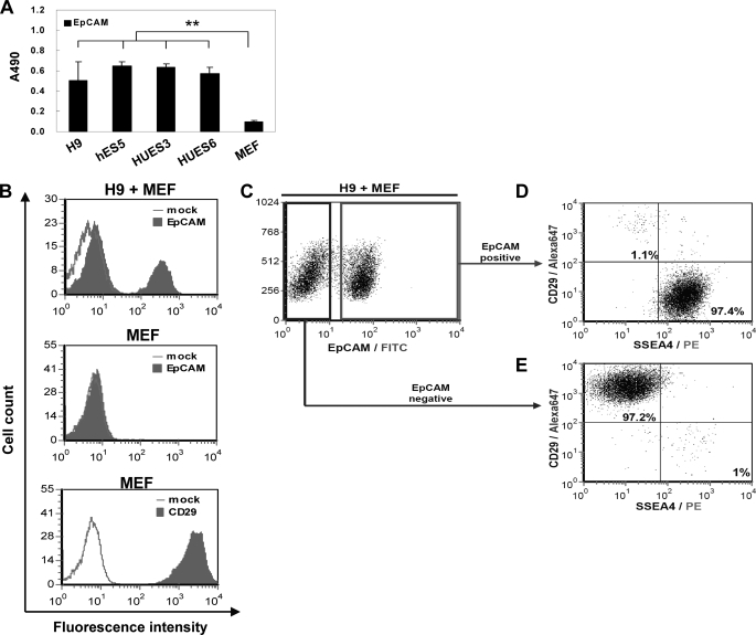
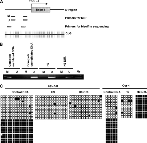
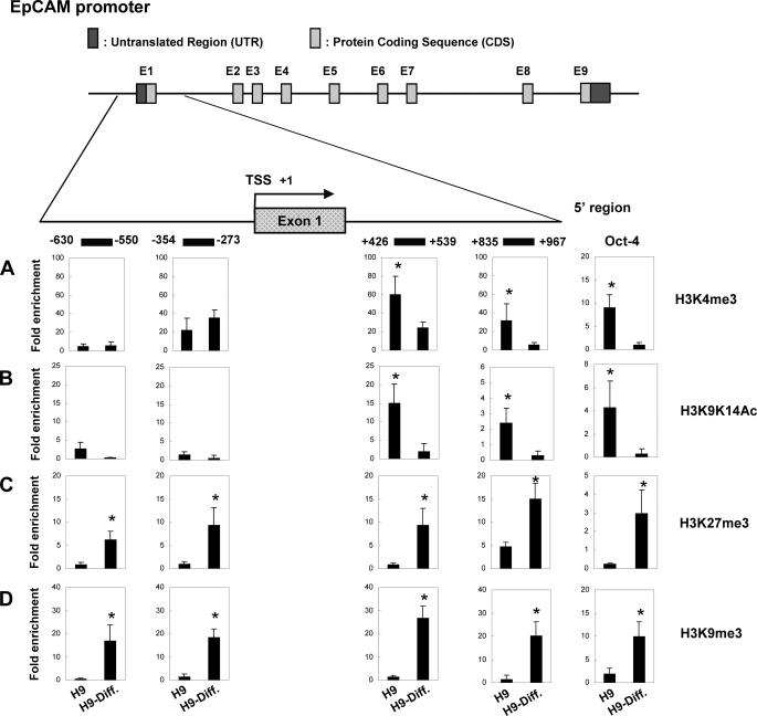
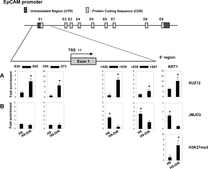
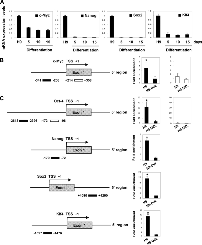
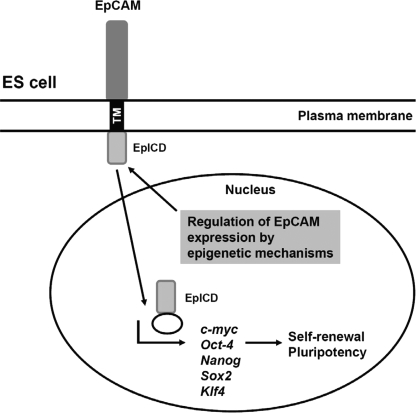
Human embryonic stem cells (hESCs) are unique pluripotent cells capable of self-renewal and differentiation into all three germ layers. To date, more cell surface markers capable of reliably identifying hESCs are needed. The epithelial cell adhesion molecule (EpCAM) is a type I transmembrane glycoprotein expressed in several progenitor cell populations and cancers. It has been used to enrich cells with tumor-initiating activity in xenograft transplantation studies. Here, we comprehensively profile the expression of EpCAM by immunofluorescence microscopy, Western blotting, and flow cytometry using an anti-EpCAM monoclonal antibody (mAb) OC98-1. We found EpCAM to be highly and selectively expressed by undifferentiated rather than differentiated hESCs. The protein and transcript level of EpCAM rapidly diminished as soon as hESC had differentiated. This silencing was closely and exclusively associated with the radical transformation of histone modification at the EpCAM promoter. Moreover, we demonstrated that the dynamic pattern of lysine 27 trimethylation of histone 3 was conferred by the interplay of SUZ12 and JMJD3, both of which were involved in maintaining hESC pluripotency. In addition, we used chromatin immunoprecipitation analysis to elucidate the direct regulation by EpCAM of several reprogramming genes, including c-MYC, OCT-4, NANOG, SOX2, and KLF4, to help maintain the undifferentiation of hESCs. Collectively, our results suggest that EpCAM might be used as a surface marker for hESC. The expression of EpCAM may be regulated by epigenetic mechanisms, and it is strongly associated with the maintenance of the undifferentiated state of hESCs.


Figures

FIGURE 1.
FIGURE 1.
EpCAM is selectively expressed by hESCs. A, dose-dependent increase in relative fluorescence intensity was associated with increasing the concentration of anti-EpCAM mAb (OC98-1) against the cell surface of undifferentiated hESCs (top; H9, hES5, and HUES6). Only basal level of EpCAM binding to 30 day-differentiated hESCs (H9-Diff., hES5-Diff., and HUES6-Diff.) can be seen in the lower panel. The differentiation of hESCs was as described under ”Experimental Procedures.“ B, expression of the EpCAM protein in undifferentiated and differentiated hESCs. Lysates from various hESCs cell lines were analyzed by Western blot analysis with the anti-EpCAM and anti-α tubulin mAb. α-Tubulin was used as an internal control. C, immunofluorescent analysis of EpCAM (i and iii) and OCT-4 protein (ii and iv) expression in undifferentiated H9 (i and ii) and differentiated H9 (iii and iv) cells. Nuclei were counterstained with 4′,6-diamidino-2-phenylindole (DAPI) (blue). H9 cells staining for OCT-4 manifested that these hESCs maintained an undifferentiated state. EpCAM expression was correlated to that of OCT-4 in undifferentiated H9 (i and ii). hESC differentiation was associated with loss of both EpCAM and OCT-4 expression (iii and iv).
FIGURE 2.
FIGURE 2.
Human ES cell differentiation was associated with loss of EpCAM. A, cell surface EpCAM expression histogram was assessed in undifferentiated H9 cells (left) and H9 cells differentiated for 5, 10, and 15 days by fluorescent flow cytometry. EpCAM or SSEA4 was a closed population, and secondary antibody only was an open population. Viable cells were gated using forward and side scatter, and the data represents cells from this population. B, Q-RT-PCR analysis of EpCAM, OCT-4, and COL3A1 transcript expression was assessed in undifferentiated H9 cells and in H9 cells differentiated for 5, 10, and 15 days as described above. GAPDH expression was used to normalize the variability in each template loading.
FIGURE 3.
FIGURE 3.
Cell surface EpCAM expression and isolation of hESCs. A, cell surface EpCAM protein expression by undifferentiated hESCs (H9, hES5, HUES3, and HUES6) and feeder MEF by ELISA using an anti-EpCAM mAb (**, p < 0.01). B, flow cytometry analysis of EpCAM on H9 hESCs co-cultured with MEF (top) and MEF alone (middle) and analysis of CD29 on MEF alone (bottom). C, analysis of cell surface expression of EpCAM on H9 cells co-cultured with MEF by fluorescent flow cytometry. D and E, double labeling of EpCAM positive (D) and EpCAM negative (E) population with anti-SSEA4 and anti-CD29 antibodies on undifferentiated H9 cells co-cultured with MEF.
FIGURE 4.
FIGURE 4.
Methylation status of EpCAM promoter regions in undifferentiated and differentiated hESCs. A, schematic representation of the EpCAM gene promoter region. Primers for MSP and bisulfite sequencing used in the study are indicated. B, MSP analysis of the EpCAM gene promoter region in undifferentiated and differentiated H9 cells. The PCR products that were methylated (M) were generated by methylation-specific primers, and those that were unmethylated (U) were generated by primers specific for unmethylated DNA. C, mapping the methylation status of the CpG islands in the promoter region of the EpCAM gene by bisulfite sequencing. Each row of squares represents a single plasmid cloned and sequenced from PCR products generated from amplification of bisulfite-treated DNA. Open squares, unmethylated cytosines; filled squares, methylated cytosines. Most CpGs in the promoter region in both undifferentiated and differentiated H9 cells were unmethylated.
FIGURE 5.
FIGURE 5.
Histone modification at the EpCAM promoter in undifferentiated and differentiated hESCs. Top, schematic representation of the EpCAM gene promoter region, which spanned positions −630 to +967 with respect to the TSS. The ChIP primers used in the study are indicated by horizontal lines. A–D, a combination of ChIP and Q-PCR analyses showing quantitative occupancy of H3K4me3 (A), H3K9K14Ac (B), H3K27me3 (C), and H3K9me3 (D) to EpCAM and OCT-4 promoter in undifferentiated and differentiated H9 cells. OCT-4 was used as a positive control for histone modification binding. Each experiment was done in triplicate (mean ± S.D.). The amount of immunoprecipitated target was quantified by real-time PCR, and the value of immunoprecipitated target was calculated as the ratio of IP DNA to the total amount of input DNA used for the immunoprecipitation (IP/input). To obtain relative -fold enrichment value, the target IP/input was further normalized to the level of a control promoter region of HBB (H3K4me3 or H3K9K14Ac) or of GAPDH (H3K27me3 or H3K9me3). In ChIP analyses, H3K4me3 and H3K9K14Ac enrichment were observed in undifferentiated H9 cells downstream of TSS, whereas H3K27me3 and H3K9me3 occupancy were detected in differentiated H9 cells both upstream and downstream of TSS (*, p < 0.05).
FIGURE 6.
FIGURE 6.
Recruitment of chromatin modifier SUZ12 and JMJD3 to EpCAM promoter in undifferentiated and differentiated hESCs. Top, schematic representation of the EpCAM promoter locus, which spanned positions −630 to +967 with respect to the TSS. The ChIP primers used in the study are indicated by horizontal lines. A and B, chromatin samples were immunoprecipitated with anti-SUZ12 antibody (A) or anti-JMJD3 antibody (B), and enrichment of the EpCAM and KRT1 promoter was quantitated by Q-PCR. KRT1 was used as a control for SUZ12/JMJD3/H3K27me3 binding. Each experiment was done in triplicate (mean ± S.D.). The value of immunoprecipitated target was calculated as the ratio of IP DNA to the total input DNA (IP/input). The target IP/input was further normalized to a control promoter region of HBB (SUZ12) or of GAPDH (JMJD3) to obtain -fold enrichment values. By ChIP measurement, the association of SUZ12 with the EpCAM promoter was elevated both upstream and downstream of TSS in differentiated H9 cells. In contrast, quantification of the intensities of JMJD3 binding was increased downstream of TSS in undifferentiated H9 cells (*, p < 0.05).
FIGURE 7.
FIGURE 7.
EpCAM regulates c-MYC, OCT-4, NANOG, SOX2, and KLF4 to help maintain stemness in hESCs. A, Q-RT-PCR analysis of mRNA expression in undifferentiated H9 cells and in H9 cells differentiated for 5, 10, and 15 days. The expression level was normalized to internal control GAPDH. B, quantitative ChIP analysis of EpCAM binding to c-MYC promoter (*, p < 0.05). C, quantitative ChIP analysis of EpCAM binding to OCT-4, NANOG, SOX2, and KLF4 promoters (*, p < 0.05). The experiment was done in triplicate (mean ± S.D.).
FIGURE 8.
FIGURE 8.
Schematic illustration of signaling pathways of EpCAM. EpCAM expression in hESCs is controlled by epigenetic regulation. The signaling of EpCAM was achieved by EpICD translocation into the nucleus, which contacted promoters of c-MYC, OCT-4, NANOG, SOX2, and KLF4 to exert its impact on maintaining the ES cell stemness condition.

Similar articles

Cited by

References

    1. Thomson J. A., Itskovitz-Eldor J., Shapiro S. S., Waknitz M. A., Swiergiel J. J., Marshall V. S., Jones J. M. (1998) Science 282, 1145–1147 - PubMed
    1. Litvinov S. V., Velders M. P., Bakker H. A., Fleuren G. J., Warnaar S. O. (1994) J. Cell Biol. 125, 437–446 - PMC - PubMed
    1. Litvinov S. V., Balzar M., Winter M. J., Bakker H. A., Briaire-de Bruijn I. H., Prins F., Fleuren G. J., Warnaar S. O. (1997) J. Cell Biol. 139, 1337–1348 - PMC - PubMed
    1. Balzar M., Winter M. J., de Boer C. J., Litvinov S. V. (1999) J. Mol. Med. 77, 699–712 - PubMed
    1. Litvinov S. V., van Driel W., van Rhijn C. M., Bakker H. A., van Krieken H., Fleuren G. J., Warnaar S. O. (1996) Am. J. Pathol. 148, 865–875 - PMC - PubMed

Publication types

MeSH terms