. 2010 Mar 19;285(12):8585-94.
doi: 10.1074/jbc.M109.090761. Epub 2010 Jan 11.

Splicing of yeast aI5beta group I intron requires SUV3 to recycle MRS1 via mitochondrial degradosome-promoted decay of excised intron ribonucleoprotein (RNP)

Affiliations

Splicing of yeast aI5beta group I intron requires SUV3 to recycle MRS1 via mitochondrial degradosome-promoted decay of excised intron ribonucleoprotein (RNP)

Edward M Turk et al. J Biol Chem. .

Abstract

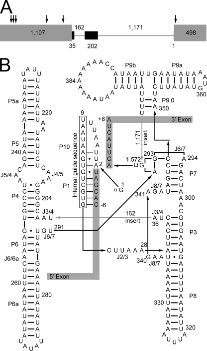
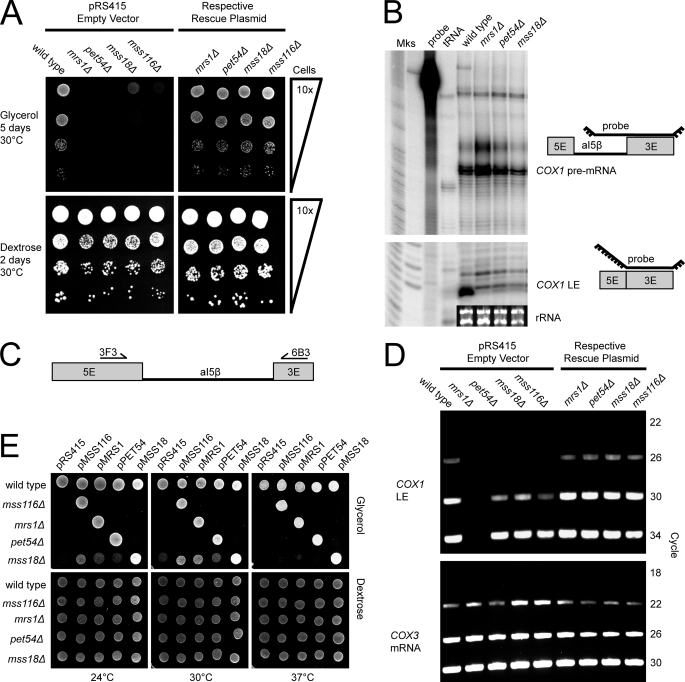
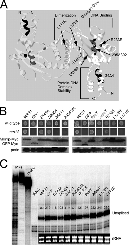
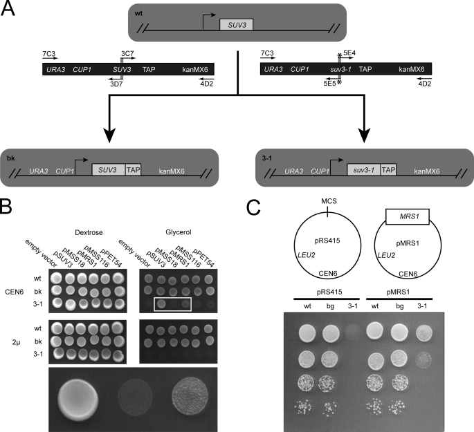
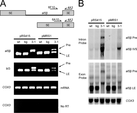
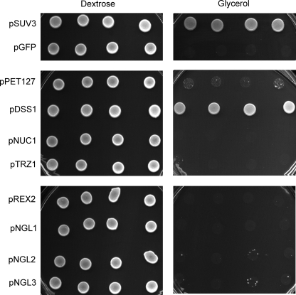
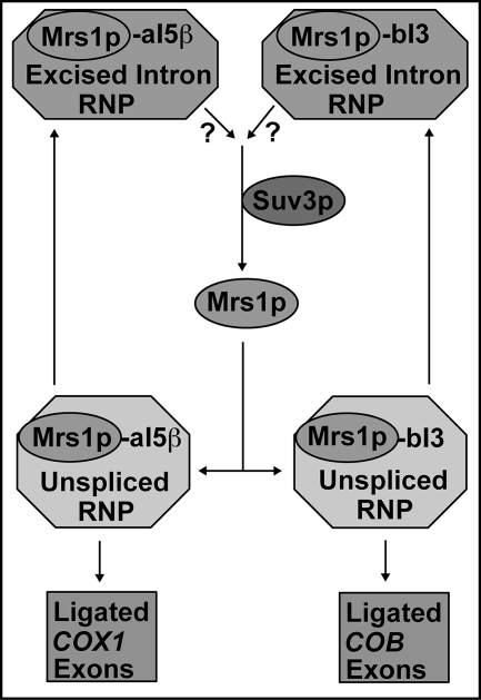
Yeast Suv3p is a member of the DEXH/D box family of RNA helicases and is a critical component of the mitochondrial degradosome, which also includes a 3' --> 5' exonuclease, Dss1p. Defects in the degradosome result in accumulation of aberrant transcripts, unprocessed transcripts, and excised group I introns. In addition, defects in SUV3 result in decreased splicing of the aI5beta and bI3 group I introns. Whereas a role for Suv3p in RNA degradation is well established, the function of Suv3p in splicing of group I introns has remained elusive. It has been particularly challenging to determine if Suv3p effects group I intron splicing through RNA degradation as part of the degradosome, or has a direct role in splicing as a chaperone, because nearly all perturbations of SUV3 or DSS1 result in loss of the mitochondrial genome. Here we utilized the suv3-1 allele, which is defective in RNA metabolism and yet maintains a stable mitochondrial genome, to investigate the role of Suv3p in splicing of the aI5beta group I intron. We provide genetic evidence that Mrs1p is a limiting cofactor for aI5beta splicing, and this evidence also suggests that Suv3p activity is required to recycle the excised aI5beta ribonucleoprotein. We also show that Suv3p acts indirectly as a component of the degradosome to promote aI5beta splicing. We present a model whereby defects in Suv3p result in accumulation of stable, excised group I intron ribonucleoproteins, which result in sequestration of Mrs1p, and a concomitant reduction in splicing of aI5beta.


Figures

FIGURE 1.
FIGURE 1.
Conserved structure of the aI5β group I intron is interrupted by two large inserts. A, COX1 pre-mRNA. All UTRs and introns, except aI5β, have been excluded for clarity. Gray rectangles represent COX1 exons, arrows show the exon junctions for the spliced out introns, black rectangles represent the conserved group I intron structure of aI5β, and inserts that disrupt the conserved structure are represented by gray lines. Length in bases for the exons, intron, and inserts are shown within, below, and above, respectively. B, predicted secondary structure. Boxed regions represent the exons, and the intronic paired regions (P) and junctions between paired regions (J) are indicated. Arrows point in the 5′ to 3′ direction to indicate linear connectivities of the nucleic acid strand. Numbering starts with the exogenous guanosine (1G), which is added during the 5′ excision reaction, and ends with the last base of the intron (1,572G), which is always a guanosine in the case of group I introns. The catalytic core consists of a paired region (P7) and a junction between two paired regions (J6/7). P7 and J6/7 are identical in sequence to the catalytic core of the Azoarcus pre-tRNA-ILE group I intron, which has been crystallized (50). Gray numbered arrows indicate the position and length of inserts that disrupt the conserved structure.
FIGURE 2.
FIGURE 2.
MRS1 is essential for aI5β splicing, while PET54, MSS18, and MSS116 are splicing enhancers. A, growth assay for mitochondrial deficiency. Deletion strains and wild type (ade2Δ) were grown on SCAG/-Leu (top) or SCAD/-Leu (bottom) as a loading control. The top right panel is a control in which each strain is rescued by a plasmid borne copy of the deleted gene. B, RNase protection assay for splicing deficiency. COX1 pre-mRNA harboring the aI5β intron (top), COX1-ligated exons (LE, bottom), Mks (markers), probe (no RNase control), tRNA (no mitochondrial RNA control), and inset is the rRNA-loading control. C, primers used for RT-PCR. Unspliced aI5β is too large to be amplified and thus only LE are detected. D, RT-PCR assay for splicing deficiency. The top panel shows COX1 LE at various cycles. The bottom panel shows amplification of intronless COX3 mRNA as a loading control. E, growth assay for functional redundancy. The top panel shows growth on glycerol, and the bottom panel is a loading control on dextrose.
FIGURE 3.
FIGURE 3.
Structural requirements of the Mrs1p protein for facilitating splicing of the aI5β intron. A, structure of the S. pombe mitochondrial four-way resolvase enzyme Ydc2p. The coordinates were downloaded from the Protein Data Bank (Accession Number: 1KCF) and visualized with the Swiss-Pdb viewer (36). One monomer of the dimer has domains labeled. Mutations introduced into the homologous Mrs1p protein are marked; the numbering system is that of Mrs1p. C-terminal truncations of 9 and 24 amino acids (9aaT, 24aaT) are mutations of Mrs1p that are not homologous to the Ydc2p protein depicted in the crystal structure. B, growth phenotypes of Myc-tagged mutant proteins expressed in the mrs1Δ and wild type (BY4741) backgrounds. Strains were grown on SCG/-Ura for 5 days at 30 °C. Western blots of total mitochondrial protein using anti-Myc or anti-porin as a loading control. C, RNase protection assay for aI5β-containing COX1 pre-mRNA. Quantification is presented above each band as a percentage of unspliced in the MRS1 lane). The probe is same as Fig. 2B, but digestion was optimized for pre-mRNA detection (see “Experimental Procedures”). Mks (markers), probe (no RNase control), tRNA (no mitochondrial RNA control), and the inset is the rRNA-loading control.
FIGURE 4.
FIGURE 4.
Genetic interaction between SUV3 and MRS1. A, scheme to make chromosomal alterations of the BY4741 wild-type yeast strain (wt), to generate the CUP1-driven SUV3 TAP-tagged background strain (bg) and an identical strain with the exception of the suv3-1 point mutation (3-1). Overlapping PCR was used to generate the URA3::CUP1::SUV3::TAP::kanMX6 product or the identical molecule with the exception of the suv3-1 point mutation. Primers are shown as arrows, and the * indicates inclusion of the suv3-1 point mutation within the primer. Templates include a plasmid with a URA3::CUP1::SUV3 cassette and a yeast strain with the SUV3-TAP-tagged allele from Open Biosystems. B, growth assay to determine if there is a genetic interaction between the suv3-1 allele and the known aI5β splicing cofactors, which are expressed from low copy (CEN6) or high-copy (2μ) vectors. 5 μl of saturated cell cultures were spotted to dextrose (SCD/-Leu) or glycerol (SCG/-Leu) plates lacking leucine and incubated at 30 °C for 7 days. The bottom row is an enlargement of the boxed section in the top row. C, dilution series growth assay on SCG/-Leu at 30 °C for 5 days to determine the relative strength of suppression of the suv3-1 glycerol growth defect by MRS1. Cells were also spotted to SCG/-Leu and SCD/-Leu control plates, which were incubated at 18, 24, 30, or 37 °C (data not shown).
FIGURE 5.
FIGURE 5.
Molecular splicing defects associated with CUP1::suv3-1::TAP can be partially rescued by overexpression of MRS1. A, analysis of aI5β and bI3 splicing by multiplex RT-PCR. The wt, bg, and 3-1 strains are the wild type, background, and suv3-1 strains described in Fig. 4. COX1 RNA was reverse-transcribed, and the cDNA amplified with the three primers shown in the scheme at the top (exons and intron are to scale). Pre, aI5β-containing COX1 pre-mRNA; LE, ligated exon. Note that the PCR conditions do not result in efficient amplification of intron-containing cDNA with 5′ and 3′ exon primers (see “Experimental Procedures”). A similar strategy, with appropriate primers, was used to analyze bI3 splicing. B, analysis of aI5β splicing by Northern blot. Total mitochondrial RNA was probed with 32P-labeled DNA oligonucleotide probes that hybridized to the aI5β intron (Intron Probe) or the exon adjacent to the 3′-end of the intron (Exon Probe) or the COX3 mRNA. IVS, free aI5β intron. The intronless COX3 mRNA was probed as a loading control.
FIGURE 6.
FIGURE 6.
Glycerol growth defect in the CUP1::suv3-1::TAP strain is rescued by overexpression of mitochondrial ribonucleases. A, CUP1::suv3-1::TAP strain was transformed with low copy vectors (pRS415) expressing GFP as a vector control or mitochondrial ribonucleases (PET127, DSS1, TRZ1, and REX2) or genes with homology to ribonucleases (NGL1 thru -3) that show mitochondrial defects when disrupted (51). Four individual transformants per vector were spotted to SCD/-Leu (left) or SCG/-Leu plates (right) and grown at 30 °C for 2 or 10 days respectively. Note that some NGL2 and 3 spots show spontaneous revertants, and these may represent suppression via mutations in the mitochondrial RNA polymerase gene as observed previously (46).
FIGURE 7.
FIGURE 7.
Proposed working model for recycling of the Mrs1p splicing cofactor by Suv3p. The excised aI5β and bI3 intron RNA is complexed with the Mrs1p protein and potentially other splicing factors (top). The Suv3p protein, acting through its degradosome function, liberates Mrs1p by degradation of the RNA (middle). Mrs1p participates in the next round of aI5β and bI3 splicing (bottom). Question marks reflect a gap in our knowledge of the degree to which Suv3p recycles Mrs1p from aI5β versus bI3.

Similar articles

Cited by

References

    1. Shadel G. S., Clayton D. A. (1993) J. Biol. Chem. 268, 16083–16086 - PubMed
    1. Cliften P. F., Jang S. H., Jaehning J. A. (2000) Mol. Cell. Biol. 20, 7013–7023 - PMC - PubMed
    1. Matsunaga M., Jaehning J. A. (2004) J. Biol. Chem. 279, 44239–44242 - PubMed
    1. Shadel G. S. (2004) Trends Genet 20, 513–519 - PubMed
    1. Amiott E. A., Jaehning J. A. (2006) Mol. Cell 22, 329–338 - PubMed

Publication types

MeSH terms