. 2010 Mar 5;285(10):7545-55.
doi: 10.1074/jbc.M109.078170. Epub 2010 Jan 11.

P2Y2 nucleotide receptors mediate metalloprotease-dependent phosphorylation of epidermal growth factor receptor and ErbB3 in human salivary gland cells

Affiliations

P2Y2 nucleotide receptors mediate metalloprotease-dependent phosphorylation of epidermal growth factor receptor and ErbB3 in human salivary gland cells

Ann M Ratchford et al. J Biol Chem. .

Abstract

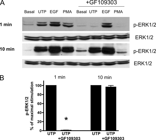
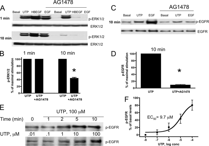
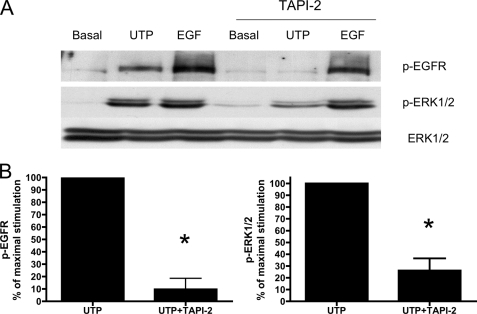
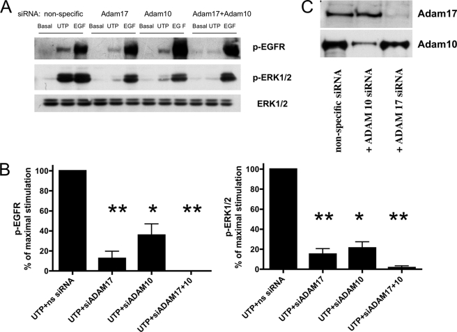
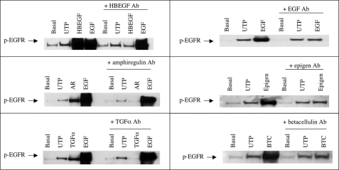
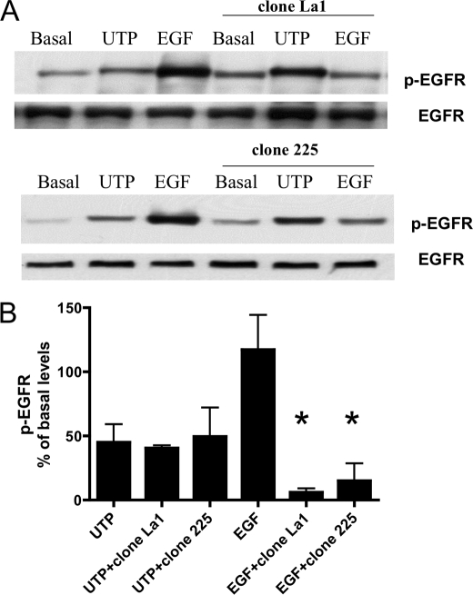
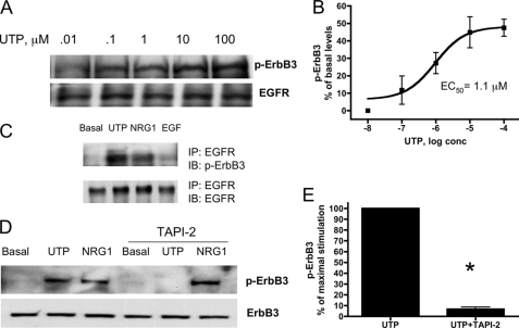
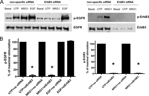
The G protein-coupled receptor P2Y(2) nucleotide receptor (P2Y(2)R) has been shown to be up-regulated in a variety of tissues in response to stress or injury. Recent studies have suggested that P2Y(2)Rs may play a role in immune responses, wound healing, and tissue regeneration via their ability to activate multiple signaling pathways, including activation of growth factor receptors. Here, we demonstrate that in human salivary gland (HSG) cells, activation of the P2Y(2)R by its agonist induces phosphorylation of ERK1/2 via two distinct mechanisms, a rapid, protein kinase C-dependent pathway and a slower and prolonged, epidermal growth factor receptor (EGFR)-dependent pathway. The EGFR-dependent stimulation of UTP-induced ERK1/2 phosphorylation in HSG cells is inhibited by the adamalysin inhibitor tumor necrosis factor-alpha protease inhibitor or by small interfering RNA that selectively silences ADAM10 and ADAM17 expression, suggesting that ADAM metalloproteases are required for P2Y(2)R-mediated activation of the EGFR. G protein-coupled receptors have been shown to promote proteolytic release of EGFR ligands; however, neutralizing antibodies to known ligands of the EGFR did not inhibit UTP-induced EGFR phosphorylation. Immunoprecipitation experiments indicated that UTP causes association of the EGFR with another member of the EGF receptor family, ErbB3. Furthermore, stimulation of HSG cells with UTP induced phosphorylation of ErbB3, and silencing of ErbB3 expression inhibited UTP-induced phosphorylation of both ErbB3 and EGFR. UTP-induced phosphorylation of ErbB3 and EGFR was also inhibited by silencing the expression of the ErbB3 ligand neuregulin 1 (NRG1). These results suggest that P2Y(2)R activation in salivary gland cells promotes the formation of EGFR/ErbB3 heterodimers and metalloprotease-dependent neuregulin 1 release, resulting in the activation of both EGFR and ErbB3.


Figures

FIGURE 1.
FIGURE 1.
UTP time- and dose-dependent phosphorylation of ERK1/2 in HSG cells. A, representative Western blots of HSG cells serum-starved for 18 h and treated with or without UTP (100 μm) for the indicated times (left top panel) with the indicated UTP concentrations for 10 min (right top panel) at 37 °C. Protein extracts were subjected to SDS-PAGE and immunoblotted for either p-ERK1/2 or ERK1/2. B, data are expressed as fold increases in ERK1/2 phosphorylation induced by UTP, as compared with untreated control, and represent the means ± S.E. of results from five experiments. conc, concentration.
FIGURE 2.
FIGURE 2.
The PKC inhibitor GF109303 attenuates rapid (1 min), but not slower and prolonged (10 min), UTP-induced ERK1/2 phosphorylation. A, representative Western blots of HSG cells serum-starved for 18 h, pretreated for 30 min with or without the PKC inhibitor, GF109303 (10 μm), and then incubated with or without UTP (100 μm), the PKC activator phorbol 12-myristate 13-acetate (PMA, 1 μm), or EGF (10 ng/ml) for 1 or 10 min at 37 °C. The protein extracts were subjected to SDS-PAGE and immunoblotted for either p-ERK1/2 or ERK1/2. B, data are expressed as percentages of maximal ERK1/2 phosphorylation induced by UTP and represent the means ± S.E. of results from five experiments, where p < 0.001 (*) is a significant difference versus ERK1/2 phosphorylation induced by UTP.
FIGURE 3.
FIGURE 3.
The EGFR tyrosine kinase inhibitor AG1478 attenuates slower and prolonged (10 min) UTP-induced p-ERK1/2 and EGFR phosphorylation but not rapid (1 min) UTP-induced ERK1/2 phosphorylation. A and C, representative Western blots of HSG cells serum-starved for 18 h, pretreated for 30 min with or without the EGFR tyrosine kinase inhibitor AG1478 (10 μm), and then incubated with or without UTP (100 μm), HBEGF (10 ng/ml), or EGF (10 ng/ml) for the indicated time at 37 °C. The protein extracts were subjected to SDS-PAGE and immunoblotted for either p-ERK1/2, p-EGFR, ERK1/2, or EGFR. E, representative Western blot of HSG cells serum-starved for 18 h and treated with or without UTP (100 μm) for the indicated times or with the indicated concentration of UTP for 10 min at 37 °C. The protein extracts were subjected to SDS-PAGE and immunoblotted for p-EGFR. B, D, and F, data are expressed as percentages of maximal ERK1/2, EGFR or basal levels of EGFR phosphorylation induced by UTP and represent the means ± S.E. of results from three experiments, where p < 0.001 (*) is a significant difference versus ERK1/2 or EGFR phosphorylation induced by UTP.
FIGURE 4.
FIGURE 4.
UTP-induced ERK1/2 and EGFR phosphorylation is not dependent on Src kinase. Representative Western blots of HSG cells transfected with SMARTpool small interfering Src RNA (100 nm) using Lipofectamine 2000, as described under “Experimental Procedures.” The cells transfected with nonspecific siRNA served as controls. Transfected cells were incubated with or without UTP (100 μm) or EGF (10 ng/ml) for the indicated time at 37 °C. The protein extracts were subjected to SDS-PAGE and immunoblotted for either p-ERK1/2, p-EGFR, ERK1/2, or Src.
FIGURE 5.
FIGURE 5.
The metalloprotease inhibitor, TAPI-2, attenuates UTP-induced phosphorylation of EGFR and ERK1/2. A, representative Western blots of HSG cells serum-starved for 18 h, pretreated for 30 min with or without TAPI-2 (10 μm), and then incubated with or without UTP (100 μm) or EGF (10 ng/ml) for 10 min at 37 °C. The protein extracts were subjected to SDS-PAGE and immunoblotted for either p-ERK1/2, p-EGFR, or ERK1/2. B, data are expressed as percentages of maximal EGFR or ERK1/2 phosphorylation induced by UTP and represent the means ± S.E. of results from four experiments, where p < 0.001 (*) is a significant difference versus EGFR or ERK1/2 phosphorylation induced by UTP.
FIGURE 6.
FIGURE 6.
UTP-induced EGFR and ERK1/2 phosphorylation is dependent on ADAM17 and ADAM10. A, representative Western blots of HSG cells transfected with SMARTpool small interfering ADAM17 (ns) RNA (100 nm), ADAM10 RNA (100 nm), or both using Lipofectamine 2000, as described under “Experimental Procedures.” The cells transfected with nonspecific (ns) siRNA served as controls. Transfected cells were incubated with or without UTP (100 μm) or EGF (10 ng/ml) for 10 min at 37 °C. The protein extracts were subjected to SDS-PAGE and immunoblotted for p-ERK1/2, p-EGFR or ERK1/2. B, data are expressed as percentages of maximal EGFR or ERK1/2 phosphorylation induced by UTP and represent the means ± S.E. of results from three experiments, where p < 0.01 (*) and p < 0.001 (**) are significant differences versus EGFR or ERK1/2 phosphorylation induced by UTP and nonspecific siRNA. C, representative Western blots of HSG cells transfected with SMARTpool small interfering ADAM17 RNA (100 nm) and/or ADAM10 RNA (100 nm) or nonspecific siRNA. The protein extracts were subjected to SDS-PAGE and immunoblotted for ADAM17 or ADAM10.
FIGURE 7.
FIGURE 7.
Neutralizing antibodies to EGFR ligands do not inhibit UTP-induced phosphorylation of the EGFR in HSG cells. Representative Western blots of HSG cells preincubated for 60 min with or without anti-HBEGF (40 μg/ml), anti-EGF (50 μg/ml), anti-amphiregulin (40 μg/ml), anti-epigen (50 μg/ml), anti-TGFα (20 μg/ml), or anti-betacellulin (20 μg/ml) antibody (Ab) and then incubated for 10 min with or without UTP (100 μm), HBEGF (20 ng/ml), EGF (10 ng/ml), amphiregulin (AR; 50 ng/ml), epigen (10 ng/ml), TGFα (3 ng/ml), or betacellulin (BTC; 100 ng/ml). The data shown are representative of results from three experiments.
FIGURE 8.
FIGURE 8.
Antibodies that block the EGFR ligand-binding domain do not inhibit UTP-induced phosphorylation of EGFR. A, representative Western blots of HSG cells serum-starved for 18 h; pretreated for 30 min with or without the anti-EGFR antibodies, clone La1, or clone 225 (10 μm), and then incubated with or without UTP (100 μm) or EGF (10 ng/ml) for 10 min at 37 °C. The protein extracts were subjected to SDS-PAGE and immunoblotted for either p-EGFR or EGFR. B, data are expressed as percentages of basal EGFR phosphorylation and represent the means ± S.E. of results from three experiments, where p < 0.001 (*) is a significant difference versus EGFR phosphorylation induced by EGF.
FIGURE 9.
FIGURE 9.
UTP induces heterodimerization of EGFR and ErbB3 in HSG cells. A, representative Western blots of HSG cells serum-starved for 18 h and treated with the indicated concentration of UTP for 10 min at 37 °C. The protein extracts were subjected to SDS-PAGE and immunoblotted for either p-ErbB3 or EGFR. B, dose response data are expressed as percentages of basal levels of ErbB3 phosphorylation and represent the means ± S.E. of results from three experiments. C, representative Western blots of HSG cells serum-starved for 18 h and then incubated with or without UTP (100 μm), NRG1 (10 ng/ml), or EGF (10 ng/ml) for 10 min at 37 °C. The cell lysates were immunoprecipitated (IP) with anti-EGFR antibody, and the immunoprecipitates were resolved by SDS-PAGE. EGFR immunoprecipitates were immunoblotted (IB) with anti-p-ErbB3 antibody or anti-EGFR antibody. D, HSG cells were serum-starved for 18 h, pretreated for 30 min with or without TAPI-2 (10 μm), and then incubated with or without UTP (100 μm) or NRG1 (10 ng/ml) for 10 min at 37 °C. The protein extracts were subjected to SDS-PAGE and immunoblotted for either p-ErbB3 or ErbB3. E, data are expressed as percentages of maximal ErbB3 phosphorylation and represent the means ± S.E. of results from three experiments, where p < 0.001 (*) is a significant difference versus ErbB3 phosphorylation induced by UTP.
FIGURE 10.
FIGURE 10.
UTP-induced phosphorylation of EGFR and ErbB3 is dependent on ErbB3 expression. A, representative Western blots of HSG cells transfected with SMARTpool small interfering ErbB3 RNA (100 nm) using Lipofectamine 2000, as described under “Experimental Procedures.” The cells transfected with nonspecific (ns) siRNA served as controls. Transfected cells were incubated with or without UTP (100 μm), NRG1 (10 ng/ml), or EGF (10 ng/ml) for 10 min at 37 °C. The protein extracts were subjected to SDS-PAGE and immunoblotted for either p-EGFR, EGFR, p-ErbB3, or ErbB3. B, data are expressed as a percentage of maximal EGFR or ErbB3 phosphorylation and represent the means ± S.E. of results from four experiments, where p < 0.001 (*) is a significant difference versus EGFR or ErbB3 phosphorylation induced by UTP or NRG1.
FIGURE 11.
FIGURE 11.
UTP-induced phosphorylation of EGFR and ErbB3 is dependent on NRG1 expression. A, representative Western blots of HSG cells transfected with SMARTpool small interfering NRG1 RNA (100 nm) using Lipofectamine 2000, as described under “Experimental Procedures.” The cells transfected with nonspecific siRNA served as controls. Transfected cells were incubated with or without UTP (100 μm), NRG1 (10 ng/ml), or EGF (10 ng/ml) for 10 min at 37 °C. The protein extracts were subjected to SDS-PAGE and immunoblotted for either p-EGFR, EGFR, p-ErbB3, ErbB3, or NRG1. B, data are expressed as percentages of maximal EGFR or ErbB3 phosphorylation and represent the means ± S.E. of results from three experiments, where p < 0.01 (*) is a significant difference versus EGFR or ErbB3 phosphorylation induced by UTP.
FIGURE 12.
FIGURE 12.
P2Y2R activation causes phosphorylation of EGFR and ErbB3 in SMG cells from wild type but not P2Y2R−/− mice. Representative Western blots of SMG cells from wt C57BL/6 and C57BL/6 P2Y2R−/− mice. SMGs were isolated, enzymatically dispersed, and cultured for 72 h, as described under “Experimental Procedures.” Then cell aggregates were serum-starved for 18 h and UTP (100 μm), EGF (10 ng/ml), or NRG1 (10 ng/ml) was added for 5 or 10 min at 37 °C. The protein extracts were subjected to SDS-PAGE and immunoblotted for p-EGFR, p-ErbB3, EGFR, and ErbB3. The data shown are representative of results from three experiments.

Similar articles

Cited by

References

    1. Hsieh M., Conti M. (2005) Trends Endocrinol. Metab. 16, 320–326 - PubMed
    1. Pierce K. L., Premont R. T., Lefkowitz R. J. (2002) Nat. Rev. Mol. Cell Biol. 3, 639–650 - PubMed
    1. Fukuhara S., Chikumi H., Gutkind J. S. (2001) Oncogene 20, 1661–1668 - PubMed
    1. Dhanasekaran N., Heasley L. E., Johnson G. L. (1995) Endocr. Rev. 16, 259–270 - PubMed
    1. van Biesen T., Luttrell L. M., Hawes B. E., Lefkowitz R. J. (1996) Endocr. Rev. 17, 698–714 - PubMed

Publication types

MeSH terms

Substances

LinkOut - more resources