. 2010 Mar 5;285(10):6952-9.
doi: 10.1074/jbc.M109.055731. Epub 2010 Jan 11.

Diacylglycerol kinase delta and protein kinase C(alpha) modulate epidermal growth factor receptor abundance and degradation through ubiquitin-specific protease 8

Affiliations

Diacylglycerol kinase delta and protein kinase C(alpha) modulate epidermal growth factor receptor abundance and degradation through ubiquitin-specific protease 8

Jinjin Cai et al. J Biol Chem. .

Abstract

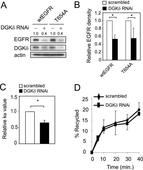
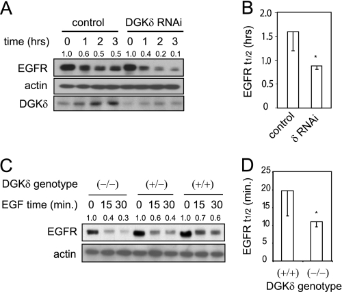
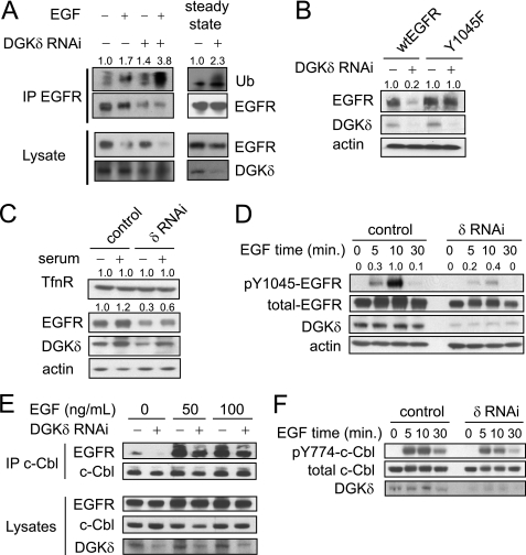
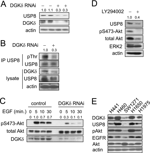
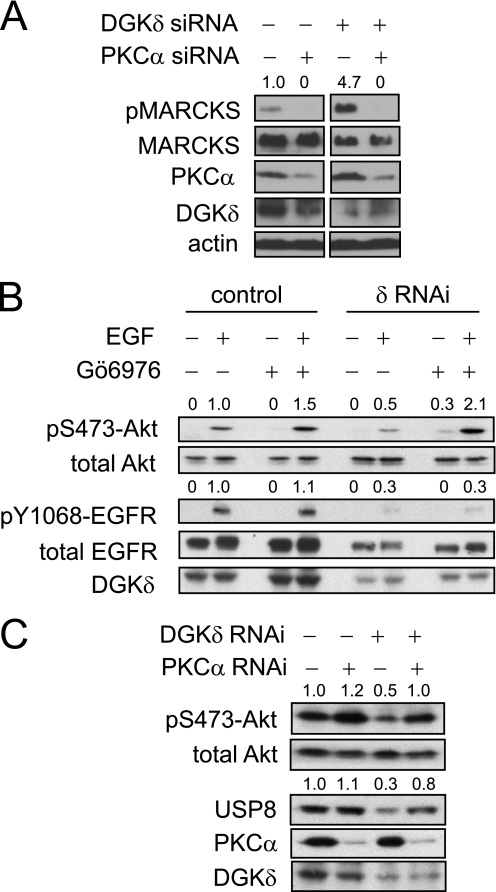
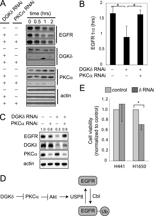
Many human epithelial cancers are characterized by abnormal activation of the epidermal growth factor receptor (EGFR), which is often caused by its excessive expression in tumor cells. The abundance of EGFR is modulated, in part, by its ubiquitination, which targets it for degradation. The components responsible for adding ubiquitin to EGFR are well characterized, but this is a reversible process, and the mechanisms that modulate the removal of ubiquitin from the EGFR are not well known. We found that de-ubiquitination of EGFR was regulated by diacylglycerol kinase delta (DGKdelta), a lipid kinase that terminates diacylglycerol signaling. In DGKdelta-deficient cells, ubiquitination of EGFR was enhanced, which attenuated the steady-state levels of EGFR and promoted its ligand-induced degradation. These effects were not caused by changes in the ubiquitinating apparatus, but instead were due to reduced expression of the de-ubiquitinase, ubiquitin-specific protease 8 (USP8). Depletion of protein kinase Calpha (PKCalpha), a target of diacylglycerol, rescued the levels of USP8 and normalized EGFR degradation in DGKdelta-deficient cells. Moreover, the effects of PKCalpha were caused by its inhibition of Akt, which stabilizes USP8. Our data indicate a novel mechanism where DGKdelta and PKCalpha modulate the levels of ubiquitinated EGFR through Akt and USP8.


Figures

FIGURE 1.
FIGURE 1.
EGFR endocytosis and recycling in DGKδ-deficient cells. A, HeLa cells were transfected with Myc-tagged wt or T654A EGFR, and then DGKδ was knocked down using siRNA. After 24 h EGFR (anti-Myc), DGKδ, and actin were detected by immunoblotting. EGFR band densities (arbitrary units) are shown above the blot. B, the band densities of wtEGFR or T654A EGFR from three experiments similar to Fig. 1A were determined and plotted. Shown are mean values with S.D. (*, p < 0.05). B, HeLa cells were transfected with scrambled control or DGKδ siRNA duplexes, and the endocytosis rate constant (ke) was determined using [125I]EGF (11). Shown are mean, relative ke values (±S.D.) from four experiments. The asterisk indicates a statistically significant difference compared with scrambled control cells (p < 0.01). C, HeLa cells were transfected with scrambled control or DGKδ siRNA duplexes, and then recycling of EGFR was measured (12). Shown are mean values (±S.D.) from three experiments.
FIGURE 2.
FIGURE 2.
Accelerated decay of EGFR in DGKδ-deficient cells. A, HeLa cells were transfected with scrambled or DGKδ siRNA duplexes, starved for 16 h, and then treated with EGF (5 ng/ml) for 0–3 h. EGFR, actin, and DGKδ were then detected in cell lysates by Western blotting. EGFR band densities (arbitrary units) are shown above the blot. Similar results were obtained when cyclohexamide (5 μm) was included to minimize new protein synthesis. B, the half-life (t½) of EGFR was measured in three experiments like that shown in A. Shown are mean values with S.D. (*, p < 0.05). C, EGFR decay in primary keratinocytes from wild-type mice or mice with heterozygous or homozygous deletion of DGKδ was measured after treatment with EGF (10 ng/ml) for the indicated times. EGFR band densities (arbitrary units) are shown above the blot. D, the half-life (t½) of EGFR was measured in three experiments like that shown in C. Shown are mean values with S.D. (*, p < 0.05).
FIGURE 3.
FIGURE 3.
Enhanced ubiquitination of EGFR in DGKδ-deficient cells is not due to an effect on c-Cbl. A, HeLa cells were transfected with scrambled or DGKδ siRNA duplexes. In the left panel, cells were starved for 16 h and then treated with EGF (10 ng/ml) for 5 min. In the right panel (steady-state), cells were grown in medium with 10% serum for 48 h. EGFR was immunoprecipitated from cell lysates (1 mg of protein for EGF-treated cells and 1.5 mg of protein for steady-state cells) and then ubiquitin (Ub), EGFR, and DGKδ were detected by Western blotting the immunoprecipitation or cell lysates. Ub band densities (arbitrary units) are shown above the blot. B, HeLa cells were transfected with Myc-tagged wild-type or Y1045F EGFR and then DGKδ was knocked down using siRNA. After 48 h EGFR (anti-Myc), DGKδ, and actin were detected by immunoblotting. EGFR band densities (arbitrary units) are shown above the blot. C, HeLa cells were transfected with scrambled or DGKδ siRNA duplexes, and then grown in complete medium or starved for 24 h. DGKδ, EGFR, transferrin receptor (TfnR), or actin were then detected in cell lysates by Western blotting. TfnR and EGFR band densities (arbitrary units) are shown above the blots. D, HeLa cells were transfected with scrambled or DGKδ siRNA duplexes, starved for 16 h, and then treated with EGF (5 ng/ml) for 0–30 min. Total EGFR, pY1045EGFR, actin, and DGKδ were detected in cell lysates by Western blotting. Band densities (arbitrary units) of pY1045EGFR normalized to total EGFR are shown above the blot. E, HeLa cells were transfected with V5-tagged c-Cbl and scrambled or DGKδ siRNA duplexes. After starvation for 16 h, the cells were exposed to 50 or 100 ng/ml EGF for 10 min, and then c-Cbl was immunoprecipitated from 1 mg of cell lysates. EGFR, c-Cbl (anti-V5), and DGKδ were detected in the precipitates and cell lysates by Western blotting. F, HeLa cells were transfected with scrambled or DGKδ siRNA duplexes, starved for 16 h, and then treated with EGF (5 ng/ml) for 0–30 min. Total c-Cbl, pY774-c-Cbl, and DGKδ were detected in cell lysates by Western blotting. All blots are representative of at least three independent experiments.
FIGURE 4.
FIGURE 4.
DGKδ deficiency reduces the levels of USP8 and attenuates Akt phosphorylation. A, in two separate experiments, HeLa cells were transfected with scrambled or DGKδ siRNA duplexes and then USP8, DGKδ, and actin were detected in cell lysates by Western blotting. USP8 band densities (arbitrary units) are shown above the blot. B, HeLa cells were transfected with V5-USP8 and then with DGKδ or control siRNA duplexes. Cells grown in medium with serum were treated with calyculin A (100 nm) for 5 min, and then lysates were harvested and V5-USP8 was immunoprecipitated. The indicated proteins were detected by Western blotting, and pThr band densities (arbitrary units) are shown above the blot. C, HeLa cells were transfected with scrambled or DGKδ siRNA duplexes, starved for 16 h, and then treated with EGF (5 ng/ml) for the indicated times. Akt, p473-Akt, and DGKδ were detected in cell lysates by Western blotting. Band densities (arbitrary units) of phosphorylated Akt are shown above the blot. D, HeLa cells growing in medium with serum were treated with 20 μm LY294002 for 24 h, and then the levels of the indicated proteins were detected in cell lysates by Western blotting. USP8 band densities (arbitrary units) are shown above the blot. E, lung cancer cell lines (noted above each lane) were grown in complete medium and harvested, and then the indicated proteins were detected by Western blotting.
FIGURE 5.
FIGURE 5.
DGKδ and PKCα modulate Akt phosphorylation and USP8 levels. A, DGKδ and/or PKCα were depleted in HeLa cells using RNAi and then the levels of phosphorylated MARCKS (pMARCKS), MARCKS, PKCα, DGKδ, and actin were determined by Western blotting. Lanes were removed from the blot for clarity. The densities of the pMARCKS bands (arbitrary units) are indicated above the blot. B, HeLa cells were transfected with scrambled or DGKδ siRNA duplexes, starved for 16 h, exposed to 250 nm Gö6976 and then to EGF (10 ng/ml) for 10 min. The levels of the indicated proteins in cell lysates were detected by Western blotting and the densities of phosphorylated Akt and EGFR (arbitrary units) are indicated above the blots. C, HeLa cells were transfected with scrambled, PKCα, and/or DGKδ siRNA duplexes, grown in medium with serum, and then the levels of the indicated proteins in cell lysates were detected by Western blotting and the densities of phosphorylated Akt and USP8 (arbitrary units) are indicated above the blots.
FIGURE 6.
FIGURE 6.
Depleting PKCα rescues the EGFR defects caused by DGKδ deficiency. A, HeLa cells were transfected with scrambled or the indicated siRNA duplexes, starved, treated with cycloheximide (5 μm, 20 min), and then exposed to EGF (5 ng/ml for 0–2 h). EGFR, DGKδ, PKCα, and actin were detected in cell lysates by Western blotting. Samples are from the same blot, but are presented to allow comparison. B, the half-life (t½) of EGFR was determined from Western blots in three independent experiments. The asterisk indicates statistical significance (p < 0.05, n = 3). C, HeLa cells were transfected with scrambled, DGKδ, and/or PKCα siRNA duplexes, grown in medium with 10% serum, and then steady-state levels of EGFR, actin, DGKδ, and PKCα were detected by Western blotting. EGFR band densities (arbitrary units) are indicated above the blot. D, our data support a model where DGKδ and PKCα regulate the activity of Akt, which in turn modulates the levels of USP8 and consequently the amount of ubiquitin attached to EGFR. E, DGKδ was knocked down using RNAi in H441 lung cancer cells (which are resistant to EGFR inhibitors) or H1650 lung cancer cells (which are sensitive) grown in medium with 2% serum. At 72 h after knockdown, cell viability was examined. Viability (mean ± S.D.) is shown normalized to cells exposed to scrambled siRNA. The asterisk indicates statistical significance (p < 0.01, n = 4). Western blotting demonstrated similar knockdown of DGKδ and EGFR in both cell lines (not shown).

Similar articles

Cited by

References

    1. Roberts R. B., Min L., Washington M. K., Olsen S. J., Settle S. H., Coffey R. J., Threadgill D. W. (2002) Proc. Natl. Acad. Sci. U.S.A. 99, 1521–1526 - PMC - PubMed
    1. Torrance C. J., Jackson P. E., Montgomery E., Kinzler K. W., Vogelstein B., Wissner A., Nunes M., Frost P., Discafani C. M. (2000) Nat. Med. 6, 1024–1028 - PubMed
    1. Buchanan F. G., Holla V., Katkuri S., Matta P., DuBois R. N. (2007) Cancer Res. 67, 9380–9388 - PubMed
    1. Sakane F., Imai S., Yamada K., Murakami T., Tsushima S., Kanoh H. (2002) J. Biol. Chem. 277, 43519–43526 - PubMed
    1. Crotty T., Cai J., Sakane F., Taketomi A., Prescott S. M., Topham M. K. (2006) Proc. Natl. Acad. Sci. U.S.A. 103, 15485–15490 - PMC - PubMed

Publication types

MeSH terms

Substances