. 2010 Mar 12;285(11):8408-21.
doi: 10.1074/jbc.M109.064386. Epub 2010 Jan 11.

Reversal of Xenopus Oct25 function by disruption of the POU domain structure

Affiliations

Reversal of Xenopus Oct25 function by disruption of the POU domain structure

Ying Cao et al. J Biol Chem. .

Abstract

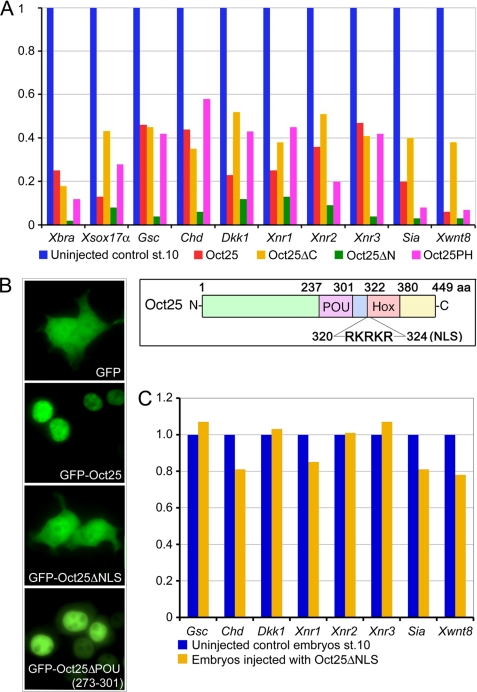
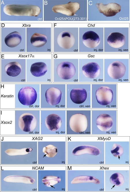
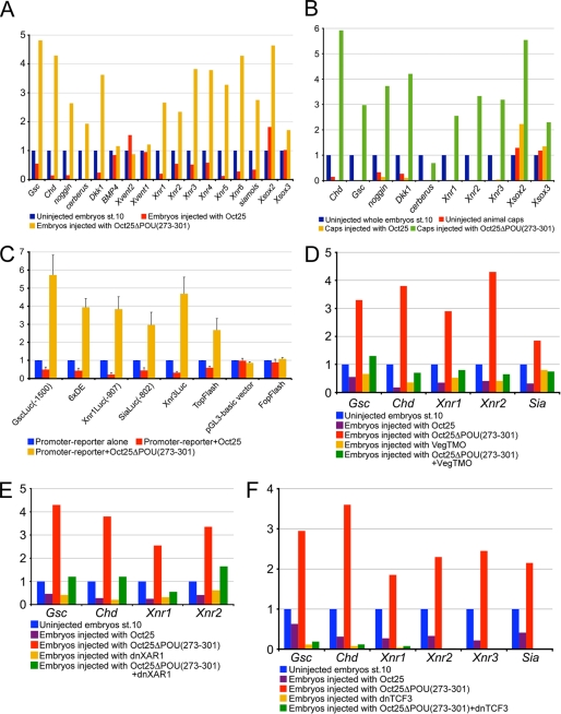
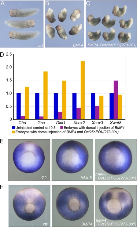
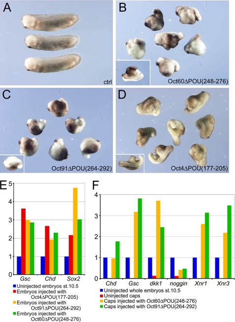
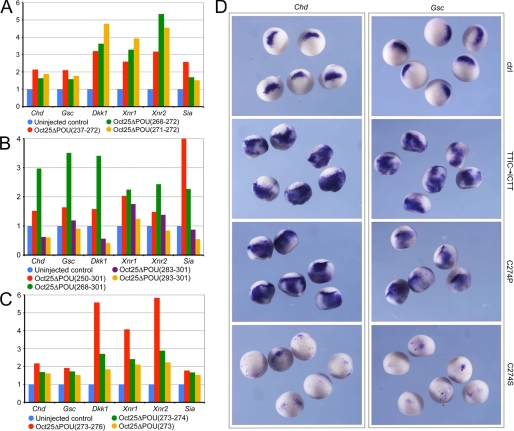
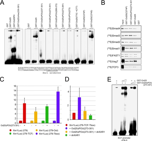
Xenopus Oct25 is a POU family subclass V (POU-V) transcription factor with a distinct domain structure. To investigate the contribution of different domains to the function of Oct25, we have performed gain of function analyses. Deletions of the N- or C-terminal regions and of the Hox domain (except its nuclear localization signal) result in mutants being indistinguishable from the wild type protein in the suppression of genes promoting germ layer formation. Deletion of the complete POU domain generates a mutant that has no effect on embryogenesis. However, disruption of the alpha-helical structures in the POU domain, even by a single amino acid mutation, causes reversal of protein function. Overexpression of such mutants leads to dorsalization of embryos and formation of secondary axial structures. The underlying mechanism is an enhanced transcription of genes coding for antagonists of the ligands for ventralizing bone morphogenetic protein and Wnt pathways. Corresponding deletion mutants of Xenopus Oct60, Oct91, or mouse Oct4 also exhibit such a dominant-negative effect. Therefore, our results reveal that the integrity of the POU domain is crucial for the function of POU-V transcription factors in the regulation of genes that promote germ layer formation.


Figures

FIGURE 1.
FIGURE 1.
Analysis of the effects of the N- and C-terminal regions and the NLS of Oct25 on gene expression. A, similar to overexpression of wild type Oct25, microinjections of RNA coding for Oct25 lacking the N-terminal region (Oct25ΔN), the C-terminal region (Oct25ΔC), or both regions (Oct25PH) lead to repression of mesodermal and endodermal inducers promoting germ layer formation. For each 400 pg of Oct25, Oct25ΔN, Oct25ΔC, or Oct25PH RNA was injected into the equatorial region of all four blastomeres at the four-cell stage. The inset shows a scheme of Oct25 structural domains. B, wild type Oct25 fused to GFP (GFP-Oct25) shows an exclusively nuclear distribution. The mutant lacking the NLS (Oct25ΔNLS) fused to GFP (GFP-Oct25ΔNLS) disperses throughout the whole cell, resembling GFP protein alone, although the mutant Oct25ΔPOU(273–301) fused to GFP localizes primarily in the nuclei. C, 1 ng of RNA of the Oct25 mutant lacking the NLS (Oct25ΔNLS) was injected into embryos. No significant effect on gene expression is observed.
FIGURE 2.
FIGURE 2.
Oct25ΔPOU(273–301) mutant reveals a strong dorsalizing activity in embryos. A and B, injection of a total of 1 ng of Oct25ΔPOU(273–301) RNA into two ventral blastomeres at the four-cell stage leads to formation of partial secondary axes (indicated with arrow in B) as compared with an uninjected control (ctrl) embryo (A). C, ventral injection of 800 pg of wild type Oct25 RNA leads to suppression of posterior structures. d–M, characterization of the resulting phenotype by whole mount in situ hybridization for selected marker genes. A total of 1 ng of RNA was injected into either two dorsal blastomeres or two ventral blastomeres at the four-cell stage as indicated for phenotype analyses. D, the pan-mesodermal marker gene Xbra is not altered significantly. E, ectopic expression of the endodermal gene Xsox17α in the marginal zone and the ectoderm. F, during gastrulation, Chordin (Chd) expression is detected in an uninjected control embryo (ctrl) at the dorsal blastopore lip. In an embryo with dorsal injection of Oct25ΔPOU(273–301) RNA, the expression domain of Chd is highly expanded (inj, dor). Moreover, Chd is also ectopically induced at the ventral side upon ventral injection (inj, ven). G, at gastrula stage, Goosecoid (Gsc) is also expressed in the dorsal lip (ctrl); dorsal injection of Oct25ΔPOU(273–301) RNA up-regulates Gsc transcription significantly (inj, dor), but ventral injection does not induce ectopic expression at the ventral side (inj, ven). H, at neurula stage, expression of epidermal keratin (keratin) is detected throughout the epidermis excluding the neural fold (ctrl, dor; ctrl, ven). In contrast, it is severely reduced in both the dorsal (inj, dor) and ventral side (inj, ven) in response to injection of Oct25ΔPOU(273–301) RNA. I, neural fold marker Xsox2 is strongly increased in response to dorsal injection of the Oct25 mutant RNA (inj, dor), and ectopic expression is observed in response to ventral injection (inj, ven) compared with uninjected embryos (ctrl, dor; ctrl, ven). J, during tailbud stage, injection of Oct25ΔPOU(273–301) RNA causes ectopic expression of XAG2, a gene that marks the most anterior structure, i.e. the cement gland. K, ectopic formation of somites is observed in injected embryos, as indicated by XMyoD expression. L, ectopic formation of neural tissue as indicated by NCAM expression. M, ectopic anterior endoderm formation, as revealed by ectopic expression of Xhex in addition to its regular expression domain. Arrows indicate ectopic gene expression.
FIGURE 3.
FIGURE 3.
Analysis of the Oct25ΔPOU(273–301) effect on gene transcription. A, injection of 1 ng of Oct25ΔPOU(273–301) RNA leads to an up-regulation of genes that induce mesoderm and endoderm and dorsalize body axis but does not generate an appreciable effect on the genes in the BMP pathway (BMP4, Xvent1, and Xvent2). B, injection of 1 ng of Oct25ΔPOU(273–301) RNA also increases the transcription of mesendoderm inducing genes and germ layer dorsalizing genes in animal caps. C, Oct25ΔPOU(273–301) stimulates luciferase reporter activity for the promoters of Gsc (GscLuc(−1500)), Xnr1 (Xnr1Luc(−907)), Xnr3 (Xnr3Luc), Sia (SiaLuc(−802)), the artificial promoter composed of six repeats of the distal element on Gsc promoter (6×DE), and the Wnt-responsive artificial promoter reporter TopFlash. For control, Oct25ΔPOU(273–301) does not have any strong effect on the pGL3-basic vector or on FopFlash, the negative control reporter for TopFlash. In each luciferase assay, 40 pg of reporter plasmid and 400 pg of RNA were injected. D–F, induction of genes by Oct25ΔPOU(273–301) is severely compromised after blocking the activities of VegT, activin/nodal, and Wnt/β-catenin signaling pathways. D, specific knockdown of VegT by 40 ng of an antisense morpholino (VegTMO) leads to reduced transcription of Gsc, Chd, Xnr1, Xnr2, and Sia. Injection of 1 ng of Oct25ΔPOU(273–301) RNA results in strong up-regulation of these genes. Up-regulation is lost when the RNA is co-injected with VegTMO. E, blocking of the nodal/activin signaling pathway by injection of 800 pg of a dominant-negative activin receptor I (dnXAR1) reduces the transcription of genes, like Gsc, Chd, Xnr1, and Xnr2, although injection of 1 ng of Oct25ΔPOU(273–301) RNA stimulates these genes. This stimulation is weakened by co-injection of dnXAR1 and Oct25ΔPOU(273–301) RNAs. F, when the Wnt/β-catenin signaling pathway is inhibited by injection of 400 pg of dominant-negative TCF3 (dnTCF3) RNA, transcription of Chd, Gsc, Xnrs, and Sia is dramatically decreased. Consequently, co-injection of 1 ng of Oct25ΔPOU(273–301) cannot stimulate the transcription of these genes anymore.
FIGURE 4.
FIGURE 4.
Oct25ΔPOU(273–301) antagonizes BMP4 activity in embryos. A, uninjected control (ctrl) embryos at tailbud stage. B, embryos show no dorsal structures upon dorsal injection of 100 pg of BMP4 RNA. C, embryos, co-injected with 100 pg of BMP4 and 1 ng of Oct25ΔPOU(273–301) RNAs together, display clear dorsal structures. D, gene expression analysis demonstrates that dorsal injection of BMP4 RNA leads to repression of dorsal genes, like Chd, Gsc, Dkk1, Xsox2, and Xsox3, and up-regulation of the ventral gene Xwnt8. This tendency is completely reversed when Oct25ΔPOU(273–301) RNA is co-injected. E, expression of the BMP-target Xvent-2 in embryos without injection (ctrl), after animal injection of hAlk-6 mRNA (1000 pg) alone, and hAlk-6 mRNA (1000 pg) in combination with Oct25ΔPOU(273–301) (500 pg). The ectopic activation on the dorsal side is not affected by Oct25ΔPOU(273–301). F, expression of Xvent-2 in embryos without injection (ctrl), after animal injection of BMP4 mRNA (1000 pg) alone, and BMP4 mRNA (1000 pg) in combination with Oct25ΔPOU(273–301) (500 pg). The ectopic activation of Xvent-2 is inhibited by Oct25ΔPOU(273–301).
FIGURE 5.
FIGURE 5.
Analysis of the effect after deletion of the region in Oct60, Oct91, and Oct4 corresponding to aa 273–301 in Oct25. A, uninjected control embryos at tailbud stage. B–D, ventral injections of 1 ng of Oct60ΔPOU(248–276) RNA (B), 1 ng of Oct91ΔPOU(264–292) RNA (C), and 1.5 ng of Oct4ΔPOU(177–205) RNA (D) lead to a dorsalized phenotype. The insets show embryos with partial double axis. E, qRT-PCRs reveal an up-regulation of dorsal genes in embryos injected with RNAs coding for Oct4ΔPOU(177–205), Oct91ΔPOU(264–292), or Oct60ΔPOU(248–276). F, qRT-PCRs of animal caps after injection with Oct91ΔPOU(264–292) or Oct60ΔPOU(248–276) RNAs also show stimulation of genes responsible for mesendoderm induction and body axis dorsalization.
FIGURE 6.
FIGURE 6.
Mutations of single amino acids cause reversal of Oct25 function and lead to an up-regulation of dorsal mesodermal genes. A, Oct25 mutants with N-terminal deletion of the POU-specific domain (aa 237–272), a few amino acids (aa 268–272), or only two amino acids (aa 271–272) cause prominent up-regulation of gene transcription in embryos. B, Oct25 mutants with depletion of a broad region of the POU-specific domain (aa 250–301 or 268–301) also have a stimulating effect on gene transcription. When a small region close to the C terminus of the POU-specific domain (aa 283–301) is truncated, this mutant exhibits either no or only rather weak stimulating activity. Up-regulation is completely lost when aa 293–301 are deleted. C, deletion of four amino acids (aa 273–276), two amino acids (aa 273–274), or a single amino acid (aa 273) leads to a strong up-regulation of gene transcription. D, whole mount in situ hybridizations demonstrate that either the change in the order of the four amino acids TTIC to ICTT (TTIC → ICTT) or the mutation of the amino acid Cys-274 to Pro (C274P) lead to an up-regulation of the transcription of Chd and Gsc. When Cys-274 is mutated to Ser, the resulting mutant represses expression of these two genes. RNA for each mutant was injected at a total of 1 ng per embryo, except for Oct25(C274S) RNA, which was injected at a total dose of 300 pg per embryo. ctrl, control.
FIGURE 7.
FIGURE 7.
DNA binding, protein interaction, and luciferase assays using Oct25 mutants. A, EMSAs (8% PAGE) show that the GST-Oct25 fusion protein binds to a canonical octamer motif (underlined) within the Xnr1 promoter. Binding is also observed for the mutant containing only the POU and Hox domains (Oct25PH). Deletion of the Hox-specific domain results in loss of DNA binding activity. GST alone was used as control. The mutants with a truncation of either the complete POU-specific domain (aa 237–301) or different regions of the POU-specific domain lose their DNA interaction capacity. DNA binding is also lost, when the amino acids TTIC are changed to ICTT (TTIC → ICTT) or Cys-274 is mutated to Pro (C274P) but is retained in the (C274S) mutant. B, GST pulldown assays show that Oct25(C274P) and Oct25(ΔPOU(273–301) still interact with Smad4, Smad2, Smad3, Smad1, FAST1, VegT, or TCF3. C, luciferase reporter activities driven by the wild type Xnr1 promoter (Xnr1Luc(−279) or the indicated deletions (15) in the absence or presence of Oct25ΔPOU(273–301). D, luciferase reporter assays driven by the −279(−TCF-Tbox) Xnr1 promoter upon co-injection with Oct25ΔPOU(−273–301) and dnXAR1. E, EMSA of the −279/−5 Xnr1 promoter fragment (6% PAGE) reveals binding of Oct25 but no interaction with Oct25ΔPOU(−273–301) GST fusion proteins.

Similar articles

Cited by

References

    1. Clements D., Friday R. V., Woodland H. R. (1999) Development 126, 4903–4911 - PubMed
    1. Hyde C. E., Old R. W. (2000) Development 127, 1221–1229 - PubMed
    1. White R. J., Sun B. I., Sive H. L., Smith J. C. (2002) Development 129, 4867–4876 - PubMed
    1. Behrens J., von Kries J. P., Kühl M., Bruhn L., Wedlich D., Grosschedl R., Birchmeier W. (1996) Nature 382, 638–642 - PubMed
    1. Wodarz A., Nusse R. (1998) Annu. Rev. Cell Dev. Biol. 14, 59–88 - PubMed

Publication types

MeSH terms

LinkOut - more resources