Comparative Study
. 2010 Sep;20(9):2198-212.
doi: 10.1093/cercor/bhp289. Epub 2010 Jan 11.

The human parahippocampal region: I. Temporal pole cytoarchitectonic and MRI correlation

Affiliations
Comparative Study

The human parahippocampal region: I. Temporal pole cytoarchitectonic and MRI correlation

X Blaizot et al. Cereb Cortex. 2010 Sep.

Abstract

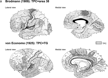
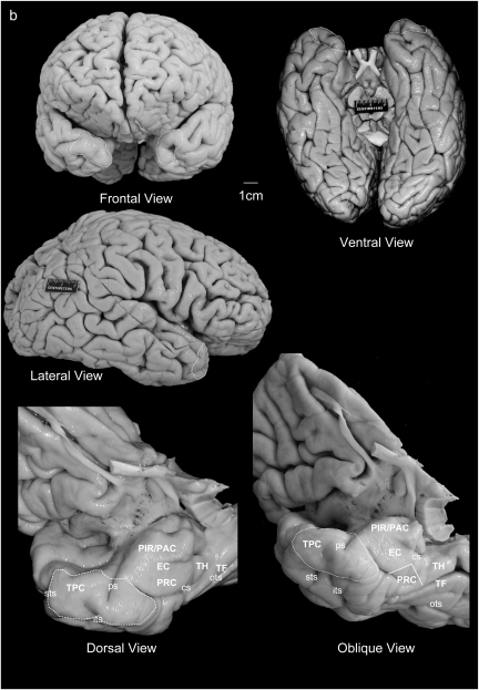
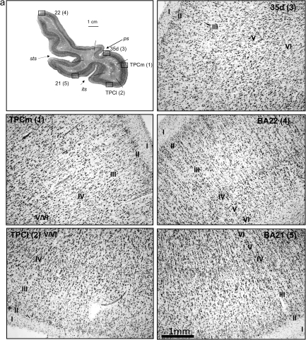
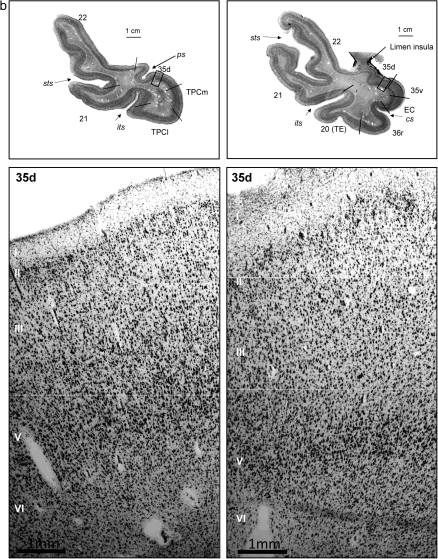
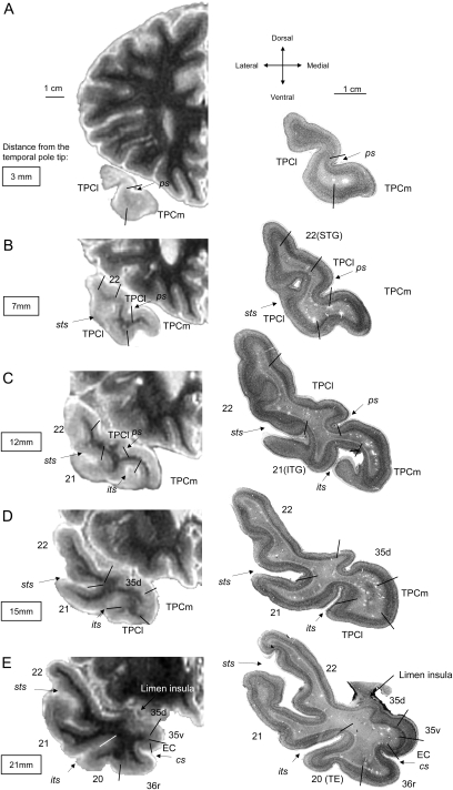
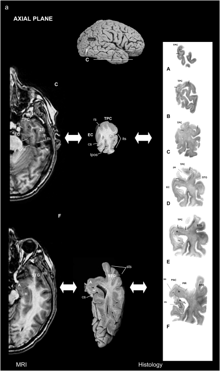
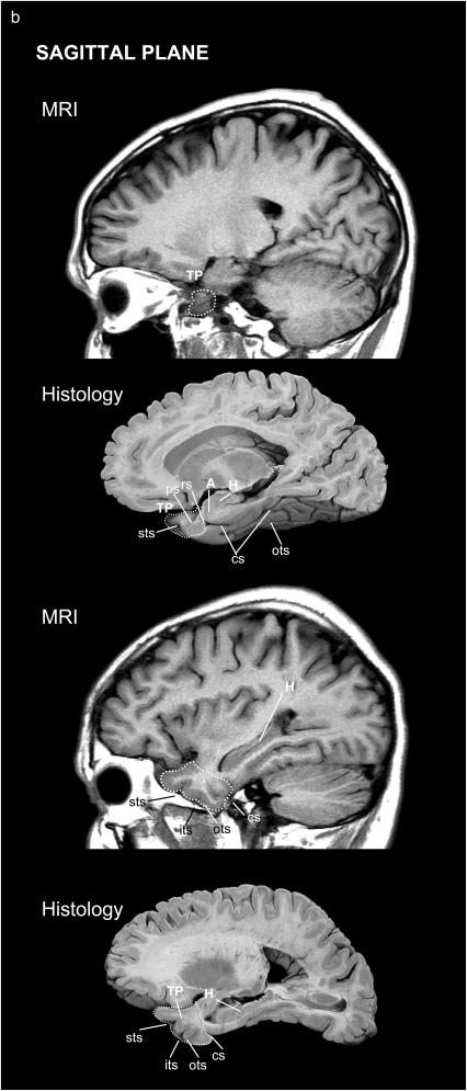
The temporal pole (TP) is the rostralmost portion of the human temporal lobe. Characteristically, it is only present in human and nonhuman primates. TP has been implicated in different cognitive functions such as emotion, attention, behavior, and memory, based on functional studies performed in healthy controls and patients with neurodegenerative diseases through its anatomical connections (amygdala, pulvinar, orbitofrontal cortex). TP was originally described as a single uniform area by Brodmann area 38, and von Economo (area TG of von Economo and Koskinas), and little information on its cytoarchitectonics is known in humans. We hypothesize that 1) TP is not a homogenous area and we aim first at fixating the precise extent and limits of temporopolar cortex (TPC) with adjacent fields and 2) its structure can be correlated with structural magnetic resonance images. We describe here the macroscopic characteristics and cytoarchitecture as two subfields, a medial and a lateral area, that constitute TPC also noticeable in 2D and 3D reconstructions. Our findings suggest that the human TP is a heterogeneous region formed exclusively by TPC for about 7 mm of the temporal tip, and that becomes progressively restricted to the medial and ventral sides of the TP. This cortical area presents topographical and structural features in common with nonhuman primates, which suggests an evolutionary development in human species.


Figures

Figure 1.
Figure 1.
(a) Location of the TPC according to Brodmann (1909) and von Economo and Koskinas (1925). (b) Frontal, ventral, and lateral aspects of the brain, with oblique views of the temporal lobe showing the whole parahippocampal gyrus including the TPC, piriform cortex, periamygdaloid, EC, PRC, and PPH cortex (areas TH and TF).
Figure 1.
Figure 1.
(a) Location of the TPC according to Brodmann (1909) and von Economo and Koskinas (1925). (b) Frontal, ventral, and lateral aspects of the brain, with oblique views of the temporal lobe showing the whole parahippocampal gyrus including the TPC, piriform cortex, periamygdaloid, EC, PRC, and PPH cortex (areas TH and TF).
Figure 2.
Figure 2.
(a) Histological appearance and cytoarchitecture of the areas constituting the TP: TPC (TPCm and TPCl), areas BA35d, BA22 (or area TE of von Economo and Koskinas), and BA21. (b) Histological appearance and cytoarchitecture of area BA35d in two sections rostrocaudally distant from 6 mm. Scale bar in low-power photomicrograph is 1 cm and 1 mm in high-magnification photomicrographs (taken with a ×5 lens).
Figure 2.
Figure 2.
(a) Histological appearance and cytoarchitecture of the areas constituting the TP: TPC (TPCm and TPCl), areas BA35d, BA22 (or area TE of von Economo and Koskinas), and BA21. (b) Histological appearance and cytoarchitecture of area BA35d in two sections rostrocaudally distant from 6 mm. Scale bar in low-power photomicrograph is 1 cm and 1 mm in high-magnification photomicrographs (taken with a ×5 lens).
Figure 3.
Figure 3.
Series of SMI-32-immunostained coronal sections, focused on layer V, of areas TPCm, TPCl, BA35d, BA22, and BA21 of the TP. Inset shows a pyramidal neuron almost completely immunostained. Scale bars are indicated.
Figure 4.
Figure 4.
T2-weighted MR images in the coronal plane of the left hemisphere of a brain embedded in agar from rostral (A) to caudal (E) levels of the TP (left). On the right-hand side of the illustration are the corresponding thionin-stained sections of the same subject (right), where cytoarchitectonically distinct fields have been depicted. Note the limited extent of TPC (labeled as TPCl or TPCm) and the heterogeneity (BA20, BA21, BA22, PRC) also present in the TP.
Figure 5.
Figure 5.
Location of the TP and surrounding areas on both MRI and corresponding histological brain sections: in the axial (a) and sagittal (b) planes.
Figure 5.
Figure 5.
Location of the TP and surrounding areas on both MRI and corresponding histological brain sections: in the axial (a) and sagittal (b) planes.
Figure 6.
Figure 6.
Left and right TPC volumes, in cubic centimeter, of the 24 subjects (left); 2D unfolded map of the parahippocampal region in one case (right).

References

    1. Arnold SE, Hyman BT, Flory J, Damasio AR, Van Hoesen GW. The topographical and neuroanatomical distribution of neurofibrillary tangles and neuritic plaques in the cerebral cortex of patients with Alzheimer's disease. Cereb Cortex. 1991;1:103–116. - PubMed
    1. Arnold SE, Hyman BT, Van Hoesen GW. Neuropathologic changes of the temporal pole in Alzheimer's disease and Pick's disease. Arch Neurol. 1994;51:145–150. - PubMed
    1. Bailey P, Bonin von G. The isocortex of man. Urbana, (IL): University of Illinois Press; 1951.
    1. Barbeau E, Sontheimer A, Joubert S, Didic M, Felician O, Tramoni E, Grimault S, Ceccaldi M, Poncet M. The human perirhinal cortex. Rev Neurol. 2004;160:401–411. - PubMed
    1. Baron JC, Chételat G, Desgranges B, Perchey G, Landeau B, de la Sayette V, Eustache F. In vivo mapping of gray matter loss with voxel-based morphometry in mild Alzheimer's disease. NeuroImage. 2001;14:298–309. - PubMed

Publication types