. 2010 Mar;78(3):1058-65.
doi: 10.1128/IAI.01141-09. Epub 2010 Jan 11.

Downregulation of PU.1 leads to decreased expression of Dectin-1 in alveolar macrophages during Pneumocystis pneumonia

Affiliations

Downregulation of PU.1 leads to decreased expression of Dectin-1 in alveolar macrophages during Pneumocystis pneumonia

Chen Zhang et al. Infect Immun. 2010 Mar.

Abstract

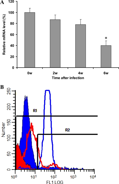
Dectin-1 is an important macrophage phagocytic receptor recognizing fungal beta-glucans. In this study, the mRNA levels of the Dectin-1 gene were found to be decreased by 61% in alveolar macrophages (AMs) from Pneumocystis-infected mice. The expression of Dectin-1 protein on the surface of these cells was also significantly decreased. By fluorescence in situ hybridization, mRNA expression levels of the transcription factor PU.1 were also found to be significantly reduced in AMs from Pneumocystis-infected mice. Electrophoretic mobility shift assay showed that PU.1 protein bound Dectin-1 gene promoter. With a luciferase reporter gene driven by the Dectin-1 gene promoter, the expression of the PU.1 gene in NIH 3T3 cells was found to enhance the luciferase activity in a dose-dependent manner. PU.1 expression knockdown by small interfering RNA (siRNA) caused a 63% decrease in Dectin-1 mRNA level and 40% decrease in protein level in AMs. Results of this study indicate that downregulation of PU.1 during Pneumocystis pneumonia leads to decreased expression of Dectin-1 in AMs.


Figures

FIG. 1.
FIG. 1.
Downregulation of Dectin-1 in AMs during PCP. (A) Real-time RT-PCR analysis of Dectin-1 mRNA in AMs. Total RNA isolated from the AMs was subjected to real-time RT-PCR analysis for Dectin-1. RPS8 was coamplified as an internal control in each assay. The mRNA expression levels of Dectin-1 in AMs from Pneumocystis-infected mice (2, 4, and 6 weeks after infection) are shown as percentages relative to that in AMs from uninfected mice. Bars represent means ± SDs for 5 or 6 mice of each group. The asterisk indicates P < 0.05 compared to control group. (B) Flow cytometry analysis of Dectin-1 protein on the surface of AMs. BAL fluid cells were stained with PE-labeled macrophage-specific antibody Mac3 and FITC-labeled anti-Dectin-1 monoclonal antibody. FL1 stands for the logarithm of FITC intensity. The blue and red lines represent anti-Dectin-1 antibody-stained AMs from uninfected and Pneumocystis-infected mice (6 weeks), respectively. The filled peaks represent the isotype control antibody-stained AMs. Gate R1 was used to include all AMs, and gate R2 was used to define Dectin-1-positive cells. Percentage of Dectin-1-positive cells was calculated by dividing the number of Dectin-1-positive cells in the R2 gate by the number of AMs in the R1 gate in each condition.
FIG. 2.
FIG. 2.
Analysis of Dectin-1 promoter. The region within 500 bp upstream from the translation initiation codon of the Dectin-1 gene was analyzed for possible transcription factor binding sites using the program Pattern Search for Transcription Factor Binding Sites (PATCH) public 1.0.
FIG. 3.
FIG. 3.
Downregulation of PU.1 in AMs during PCP. (A) Real-time RT-PCR analysis of PU.1 mRNA in AMs. Total RNA isolated from the AMs was subjected to real-time RT-PCR analysis for PU.1. RPS8 was coamplified as an internal control in each assay. The mRNA expression levels of PU.1 in AMs from Pneumocystis-infected mice (2, 4, and 6 weeks after infection) are shown as percentages relative to those in AMs from uninfected mice. Bars represent means ± SDs for 5 or 6 mice of each group. The asterisks indicate P < 0.05 compared to control group. (B) AMs from mice without or with PCP for 6 weeks were permeabilized and reacted with FITC-labeled antisense PU.1 probe. FITC-labeled sense probe was used as the negative control. PU.1 mRNA levels were examined under a fluorescence microscope at a magnification of ×400. DAPI, 4′,6-diamidino-2-phenylindole. (C) Equal amounts of total protein from mice with or without PCP were electrophoresed on a 10% SDS-polyacrylamide gel. Protein concentration of PU.1 was determined by Western blotting using anti-PU.1 antibody. GAPDH was used as an internal loading standard. Data presented are representative of 5 or 6 mice per group.
FIG. 4.
FIG. 4.
Binding of PU.1 to Dectin-1 promoter. EMSA was performed using the two oligonucleotide probes containing the putative PU.1 binding sites in Dectin-1 promoter and a recombinant PU.1 protein. The oligonucleotide containing a known PU.1-binding site was used as the positive control. For each reaction, a negative control with addition of an unrelated protein, EBNA, instead of PU.1 protein was included. Cold competition for each probe was done by adding unlabeled oligonucleotides with the known PU.1-binding site to the reaction mixture.
FIG. 5.
FIG. 5.
Enhancement of the promoter activity of the Dectin-1 gene by PU.1. The plasmid pGL-Dectin1 containing the luciferase reporter gene driven by the Dectin-1 promoter containing two putative PU.1-binding sites (black bar) (A), together with the PU.1-expressing plasmid pFLAG-PU.1 at different doses (0.1, 0.25, and 0.5 μg), was introduced into NIH 3T3 cells. Dectin-1 promoter activity was determined by measuring luciferase activity in the cell lysates (B). Bars represent SDs of three independent assays. The asterisks indicate P < 0.05 compared to the control group.
FIG. 6.
FIG. 6.
Effect of PU.1 siRNA on the expression of Dectin-1. Normal AMs (5 × 10⁵) were transfected with PU.1 siRNA or control siRNA. Dectin-1 gene expression was examined at the mRNA level by real-time PCR (A) and at the protein level by Western blotting (B). Similar results were obtained in three independent experiments. The asterisk indicates P < 0.05 compared to control group.
FIG. 7.
FIG. 7.
Effects of PU.1 or Dectin-1 knockdown on phagocytic activity of macrophages. RAW 264.7 cells (5 × 10⁵) were transfected with PU.1 siRNA, Dectin-1 siRNA, or control siRNA. The phagocytosis assay was performed by incubating transfected cells with 5 × 10⁶ FITC-labeled zymosan particles for 1 h. The cells were examined under a fluorescence microscope at a magnification of ×400 and evaluated by cell association index (CAI) representing the number of FITC-labeled zymosan particles bound to or phagocytosed per 100 macrophages. Bars represent means ± SDs for each condition in 500 cells on two separate slides. The asterisks indicate P < 0.05 compared to control group.

References

    1. Bonfield, T. L., B. Raychaudhuri, A. Malur, S. Abraham, B. C. Trapnell, M. S. Kavuru, and M. J. Thomassen. 2003. PU.1 regulation of human alveolar macrophage differentiation requires granulocyte-macrophage colony-stimulating factor. Am. J. Physiol. Lung Cell. Mol. Physiol. 285:L1132-L1136. - PubMed
    1. Chang, H. C., S. Zhang, V. T. Thieu, R. B. Slee, H. A. Bruns, R. N. Laribee, M. J. Klemsz, and M. H. Kaplan. 2005. PU.1 expression delineates heterogeneity in primary Th2 cells. Immunity 22:693-703. - PubMed
    1. DeKoter, R. P., and H. Singh. 2000. Regulation of B lymphocyte and macrophage development by graded expression of PU.1. Science 288:1439-1441. - PubMed
    1. del Pilar Jimenez, A. M., S. Viriyakosol, L. Walls, S. K. Datta, T. Kirkland, S. E. Heinsbroek, G. Brown, and J. Fierer. 2008. Susceptibility to Coccidioides species in C57BL/6 mice is associated with expression of a truncated splice variant of Dectin-1 (Clec7a). Genes Immun. 9:338-348. - PMC - PubMed
    1. De Stefano, J. A., M. T. Cushion, R. G. Sleight, and P. D. Walzer. 1990. Analysis of Pneumocystis carinii cyst wall. I. Evidence for an outer surface membrane. J. Protozool. 37:428-435. - PubMed

Publication types

MeSH terms

LinkOut - more resources