. 2010 Mar;30(6):1368-81.
doi: 10.1128/MCB.00741-09. Epub 2010 Jan 11.

Protein phosphatase 6 interacts with the DNA-dependent protein kinase catalytic subunit and dephosphorylates gamma-H2AX

Affiliations

Protein phosphatase 6 interacts with the DNA-dependent protein kinase catalytic subunit and dephosphorylates gamma-H2AX

Pauline Douglas et al. Mol Cell Biol. 2010 Mar.

Abstract

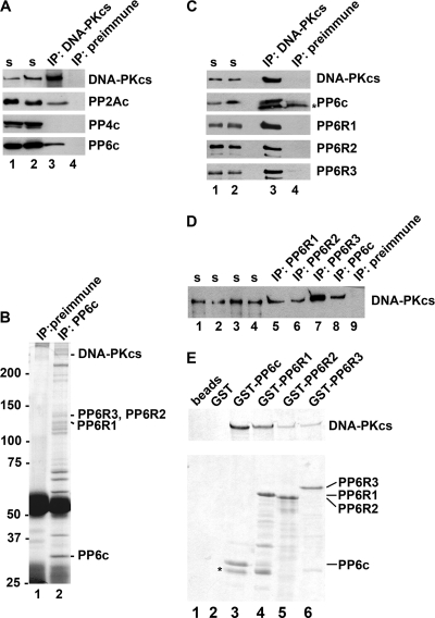
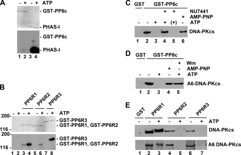
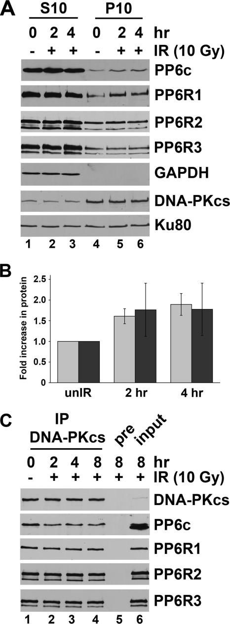
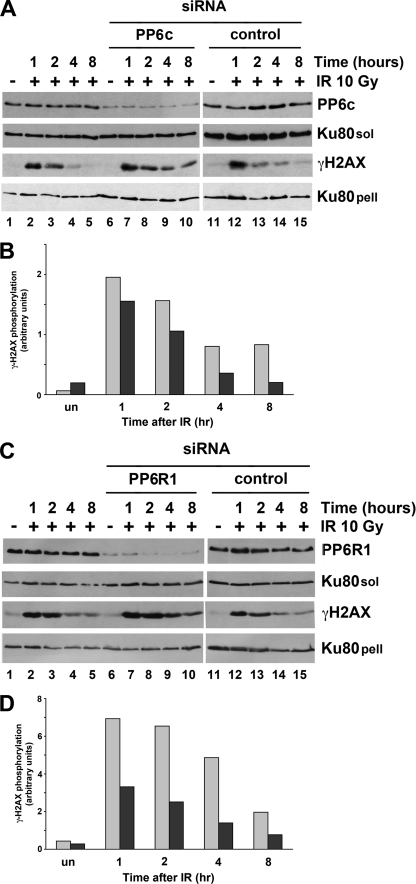
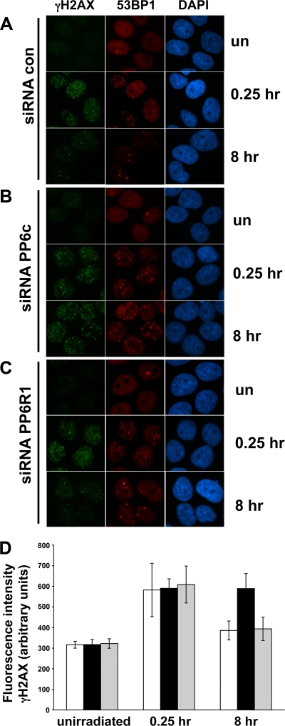
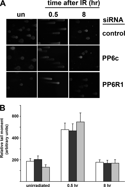
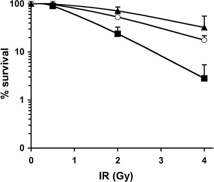
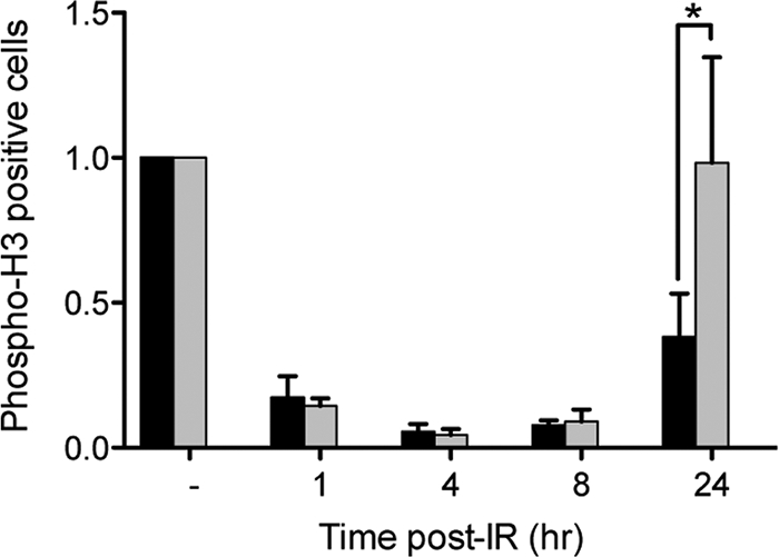
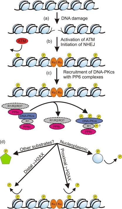
The catalytic subunit of the DNA-dependent protein kinase (DNA-PKcs) plays a major role in the repair of DNA double-strand breaks (DSBs) by nonhomologous end joining (NHEJ). We have previously shown that DNA-PKcs is autophosphorylated in response to ionizing radiation (IR) and that dephosphorylation by a protein phosphatase 2A (PP2A)-like protein phosphatase (PP2A, PP4, or PP6) regulates the protein kinase activity of DNA-PKcs. Here we report that DNA-PKcs interacts with the catalytic subunits of PP6 (PP6c) and PP2A (PP2Ac), as well as with the PP6 regulatory subunits PP6R1, PP6R2, and PP6R3. Consistent with a role in the DNA damage response, silencing of PP6c by small interfering RNA (siRNA) induced sensitivity to IR and delayed release from the G(2)/M checkpoint. Furthermore, siRNA silencing of either PP6c or PP6R1 led to sustained phosphorylation of histone H2AX on serine 139 (gamma-H2AX) after IR. In contrast, silencing of PP6c did not affect the autophosphorylation of DNA-PKcs on serine 2056 or that of the ataxia-telangiectasia mutated (ATM) protein on serine 1981. We propose that a novel function of DNA-PKcs is to recruit PP6 to sites of DNA damage and that PP6 contributes to the dephosphorylation of gamma-H2AX, the dissolution of IR-induced foci, and release from the G(2)/M checkpoint in vivo.


Figures

FIG. 1.
FIG. 1.
DNA-PKcs interacts with the catalytic and regulatory subunits of PP6. (A) DNA-PKcs was immunoprecipitated from HEK293 cells, and immunoblots were probed with antibodies to DNA-PKcs, PP2Ac, PP4c, or PP6c as indicated. Lanes 1 and 2 contained the supernatants (s) from extracts immunoprecipitated for DNA-PKcs or preimmune serum, respectively. Lanes 3 and 4 contained immunoprecipitates (IP) for DNA-PKcs or preimmune serum as indicated. (B) PP6c was immunoprecipitated from HeLa cells, and samples were analyzed by SDS-PAGE with Coomassie blue staining. PP6c-interacting proteins were identified by mass spectrometry. Indicated are bands identified as DNA-PKcs, PP6c, PP6R1, PP6R2, and PP6R3. Lane 1, preimmune serum; lane 2, PP6c serum. Positions of molecular weight markers (in thousands) are indicated on the left. (C) DNA-PKcs was immunoprecipitated from HEK293 cells as in the experiment for which results are shown in panel A. Immunoblots were probed with antibodies to DNA-PKcs, PP6c, PP6R1, PP6R2, and PP6R3, as indicated. Lanes 1 and 2, supernatants; lane 3, immunoprecipitates with antisera to DNA-PKcs; lane 4, immunoprecipitates with preimmune serum. The asterisk indicates a nonspecific, cross-reacting band in lanes 3 and 4. (D) PP6c, PP6R1, PP6R2, and PP6R3 were immunoprecipitated from HEK293 cells as in the experiments for which results are shown in panels A and C, and immunoblots were probed for DNA-PKcs. (E) PP6c, PP6R1, PP6R2, and PP6R3 were expressed in bacteria with N-terminal GST tags, immobilized on glutathione-Sepharose beads, and incubated with 5 μg purified DNA-PKcs protein (in the absence of Ku). Beads were washed as described in Materials and Methods, and proteins were analyzed by SDS-PAGE, followed by immunoblotting with an antibody to DNA-PKcs (upper panel). Lane 1, DNA-PKcs with beads alone; lane 2, DNA-PKcs plus GST-bound beads; lanes 3 to 6, DNA-PKcs plus GST-PP6c, PP6R1, PP6R2, or PP6R3 as indicated. The SDS-PAGE gel was stained with Coomassie blue to show protein loading (lower panel). The positions of migration of PP6 subunits are shown on the right. The asterisk in lane 3 represents a contaminating band.
FIG. 2.
FIG. 2.
DNA-PK-mediated phosphorylation regulates DNA-PK-PP6 interactions in vitro. (A) GST-PP6c (lanes 1 and 2) or PHAS-I (lanes 3 and 4) was incubated with DNA-PKcs and the Ku heterodimer, either in the absence of ATP (−) (lanes 1 and 3) or in the presence of [γ-32P]ATP (+) (lanes 2 and 4), under standard kinase assay conditions as described in Materials and Methods. After a 30-min incubation at 37°C, proteins were boiled in SDS sample buffer and resolved on a 12.5% SDS-PAGE gel. The gel was stained with Coomassie blue, destained, dried, and exposed to X-ray film. (Top) Coomassie-stained gel; (bottom) the corresponding autoradiograph. (B) GST, GST-PP6R1, GST-PP6R2, and GST-PP6R3 were phosphorylated by DNA-PK (DNA-PKcs plus Ku heterodimer) as described for panel A. Lanes 1 and 2, GST alone; lanes 3 and 4, GST-PP6R1; lanes 5 and 6, GST-PP6R2; lanes 7 and 8, GST-PP6R3. (Top) Coomassie-stained gel; (bottom) the corresponding autoradiograph. (C) DNA-PKcs was autophosphorylated in the presence of Ku as described in Materials and Methods and was incubated with GST-PP6c bound to glutathione beads. Beads were washed, and bound proteins were analyzed by SDS-PAGE and immunoblotted with an antibody to DNA-PKcs. Lane 1, GST bound to beads; lane 2, GST-PP6c incubated with DNA-PK (DNA-PKcs plus Ku) in a mock autophosphorylation reaction without ATP; lane 3, GST-PP6c incubated with DNA-PK in an autophosphorylation reaction with ATP; lane 4, GST-PP6c incubated with DNA-PK in the presence of the inhibitor NU7441 for 5 min before the addition of ATP; lane 5, GST-PP6c incubated with DNA-PK but with the addition of NU7441 after a 30-min incubation with ATP; lane 6, GST-PP6c incubated with DNA-PK in autophosphorylation reaction mixtures that contained the nonhydrolyzable ATP analogue AMP-PNP. (D) A6-DNA-PKcs was autophosphorylated in the presence of Ku as described in Materials and Methods and was incubated with GST-PP6c bound to glutathione beads. Beads were washed as described above and were immunoblotted for DNA-PKcs. Lanes 1 to 3 were as described for panel C. Lane 4 was an autophosphorylation reaction mixture containing A6-DNA-PKcs and AMP-PNP. In lane 5, reaction mixtures containing A6-DNA-PKcs were preincubated with wortmannin (Wm) before the addition of ATP. GST pulldown assays were performed as for panel C. (E) DNA-PKcs (top) or A6-DNA-PKcs (bottom) was autophosphorylated in the presence of Ku, either in the absence or in the presence of ATP as described above, and was then incubated with GST-PP6R1, GST-PP6R2, or GST-PP6R3 as for panel B. Lane 1, GST bound to beads; lanes 2 and 3, GST-PP6R1; lanes 4 and 5, GST-PP6R2; lanes 6 and 7, GST-PP6R3. ATP was present in the autophosphorylation reaction mixtures for the samples in lanes 3, 5, and 7. GST pulldown assays were performed as for panel C.
FIG. 3.
FIG. 3.
The catalytic and regulatory subunits of PP6 subunit localize to the nucleus, and their association with DNA-PKcs does not change after IR. (A) HEK293 cells were either left untreated (−) or treated with 10 Gy IR (+) and were left to recover for the times indicated. Cells were fractionated into cytoplasmic (S10) (lanes 1 to 3) or nuclear (P10) (lanes 4 to 6) extracts as described in Materials and Methods. Fifty micrograms of protein was run on SDS-PAGE gels and probed for PP6c, PP6R1, PP6R2, PP6R3, DNA-PKcs, GAPDH, or Ku80 as indicated. (B) P10 extracts were prepared and analyzed as for panel A. Levels of PP6c (gray bars) and PP6R1 (black bars) were normalized to nuclear Ku80 protein levels (set at 1). unIR, unirradiated. (C) DNA-PKcs was immunoprecipitated from detergent lysates from either unirradiated cells (lane 1) or cells that had been irradiated with 10 Gy and harvested 2, 4, or 8 h later as indicated (lanes 2 to 4). Immunoprecipitates were run on SDS-PAGE gels and were immunoblotted for DNA-PKcs, PP6c, PP6R1, PP6R2, or PP6R3 as indicated. Lane 5, preimmune sera (control); lane 6, input (representing 25 μg of total protein from the whole-cell extract). Note that longer exposures showed the presence of DNA-PKcs in the input lane (lane 6).
FIG. 4.
FIG. 4.
Knockdown of PP6c or PP6R1 induces sustained phosphorylation of γ-H2AX in irradiated cells. (A) HeLa cells either were left untransfected (lanes 1 to 5) or were transfected with a siRNA to either PP6c (lanes 6 to 10) or a scrambled RNA control (lanes 11 to 15). Where indicated (+), cells were irradiated with 10 Gy and were collected at the times indicated after IR. The minus sign (−) above lanes 1, 6, and 11 indicates unirradiated cells. The soluble fraction of detergent extracts was analyzed by SDS-PAGE and immunoblotted with antibodies to PP6c or Ku80 (Ku80sol) as indicated. The pellet remaining after detergent lysis was resuspended in SDS as described in Materials and Methods and was analyzed for γ-H2AX phosphorylation or Ku80 (Ku80pell) as indicated. Extracts were also probed for the expression of other protein phosphatase catalytic and regulatory subunits (shown in Fig. S4 in the supplemental material). (B) The signal intensity of γ-H2AX was normalized to that of Ku80, and the relative phosphorylation of H2AX in arbitrary units is plotted. Gray bars represent extracts from cells transfected with a siRNA to PP6c; black bars represent extracts from cells transfected with a scrambled control siRNA. (C) Cells were transfected with a siRNA to PP6R1, and extracts were generated and analyzed exactly as for panel A. Further controls are shown in Fig. S6 in the supplemental material. (D) Quantitation of γ-H2AX phosphorylation as in panel B. Gray bars represent extracts from cells transfected with a siRNA to PP6R1; black bars represent extracts from cells transfected with a scrambled control siRNA.
FIG. 5.
FIG. 5.
Knockdown of PP6c causes sustained H2AX and 53BP1 focus formation in irradiated cells. (A to C) HeLa cells were transfected either with a control siRNA (A) or with a siRNA to PP6c (B) or PP6R1 (C). After 72 h, cells were irradiated (2 Gy); then they were stained for γ-H2AX and 53BP1 focus formation at either 0.25 or 8 h post-IR. un, transfected but unirradiated cells; 0.25 hr, cells fixed and analyzed 0.25 h post-IR; 8 hr, cells fixed and analyzed 8 h post-IR. Green, γ-H2AX foci; red, 53BP1 foci; blue, DAPI staining. (D) Total nuclear intensity was determined for cells stained for γ-H2AX. In each case (control, siRNA to PP6c, or siRNA to PP6R1), the intensity at 0.25 and 8 h was compared with that of unirradiated cells. Open bars, scrambled siRNA control; filled bars, siRNA to PP6c; shaded bars, siRNA to PP6R1.
FIG. 6.
FIG. 6.
Depletion of PP6c or PP6R1 does not induce DNA damage. (A) HeLa cells were transfected with a siRNA to PP6c or to PP6R1 or with a scrambled control, as indicated on the right. Cells were irradiated with 20 Gy IR and were collected at the indicated times for neutral comet assays. A representative image of cells under each condition is presented. un, unirradiated cells. (B) Quantification of the tail lengths from the experiment for which results are shown in panel A. The tail length for each condition was calculated from a minimum of 100 cells for each data point. Open bars, scrambled siRNA control; black bars, siRNA to PP6c; gray bars, siRNA to PP6R1.
FIG. 7.
FIG. 7.
Knockdown of PP6c induces radiation sensitivity. HeLa cells were transfected either with a siRNA to PP6c or to PP6R1 or with a scrambled control. Seventy-two hours after transfection, cells were either left unirradiated or irradiated with 0.5, 2, or 4 Gy and were assayed as described in Materials and Methods. Open circles, scrambled control siRNA; triangles, siRNA to PP6R1; squares, siRNA to PP6c. Only positive error bars are shown. Results are representative of three independent experiments.
FIG. 8.
FIG. 8.
PP6 promotes recovery from the G2/M checkpoint. U2OS cells were transfected either with a siRNA against PP6c (black bars) or with a scrambled control (gray bars), they were collected at various time points after either mock IR (−) or irradiation with 3 Gy, and the relative level of mitotic cells was determined by flow cytometry using an antibody against phosphohistone H3. Statistical analysis was performed using two-way analysis of variance with Bonferroni's posttest. The asterisk indicates a P value of <0.05.
FIG. 9.
FIG. 9.
Model for the role of DNA-PKcs and PP6 in the regulation of γ-H2AX phosphorylation. (a) DNA-damaging agents, such as IR, induce the formation of DSBs in chromatin. (b) DSBs activate DNA damage response pathways, leading to activation of the ATM protein kinase and the initiation of NHEJ. Once activated, ATM phosphorylates nucleosomal H2AX on serine 139 (indicated by P in yellow circles) to form γ-H2AX. In the first step of NHEJ, the Ku70/80 heterodimer (Ku, light orange) binds to either side of the DSB, recruiting DNA-PKcs (blue), which also contributes to γ-H2AX phosphorylation. (c) In the absence of IR, DNA-PKcs interacts directly with PP6c (pink oval with solid borders), PP6R1 (gray oval with solid borders), and/or PP6R2 and PP6R3 (not shown). PP6c also interacts with any one of its three regulatory subunits (PP6R1, PP6R2, or PP6R3) (gray ovals with dashed borders). Similarly, the DNA-PKcs-PP6R1 complex interacts with PP6c (pink oval with dashed borders) through direct interaction between PP6c and PP6R1. In response to DSB formation, DNA-PKcs-PP6 complexes are recruited to Ku, where DNA-PKcs undergoes autophosphorylation and release from the DSB. Autophosphorylation of DNA-PKcs leads to dissociation of PP6c (with its associated regulatory subunits). DNA-PKcs-PP6R2 or DNA-PKcs-PP6R3 complexes also dissociate in response to autophosphorylation (not shown). PP6R1 (with its associated PP6c subunit) remains bound to autophosphorylated DNA-PKcs. PP6R1 is phosphorylated in vitro by DNA-PKcs (yellow circles with P), which may contribute to the regulation of the DNA-PKcs-PP6R1 complex. (d) The various DNA-PKcs-PP6 complexes contribute to the dephosphorylation of γ-H2AX at different sites within the nucleus; for example, different combinations of the DNA-PKcs-PP6 complex may dephosphorylate γ-H2AX that is either distal or proximal to the DSB, γ-H2AX that is present in the nucleoplasm rather than bound to chromatin, or γ-H2AX that exists in the proximity of heterochromatin (not shown). PP6 complexes may also dephosphorylate other proteins present at or in the vicinity of foci (green pentagon).

Similar articles

Cited by

References

    1. Achari, Y., and S. P. Lees-Miller. 2000. Detection of DNA-dependent protein kinase in extracts from human and rodent cells. Methods Mol. Biol. 99:85-97. - PubMed
    1. Ahnesorg, P., P. Smith, and S. P. Jackson. 2006. XLF interacts with the XRCC4-DNA ligase IV complex to promote DNA nonhomologous end-joining. Cell 124:301-313. - PubMed
    1. Allalunis-Turner, M. J., P. K. Zia, G. M. Barron, R. Mirzayans, and R. S. Day III. 1995. Radiation-induced DNA damage and repair in cells of a radiosensitive human malignant glioma cell line. Radiat. Res. 144:288-293. - PubMed
    1. Bakkenist, C. J., and M. B. Kastan. 2003. DNA damage activates ATM through intermolecular autophosphorylation and dimer dissociation. Nature 421:499-506. - PubMed
    1. Beausoleil, S. A., M. Jedrychowski, D. Schwartz, J. E. Elias, J. Villen, J. Li, M. A. Cohn, L. C. Cantley, and S. P. Gygi. 2004. Large-scale characterization of HeLa cell nuclear phosphoproteins. Proc. Natl. Acad. Sci. U. S. A. 101:12130-12135. - PMC - PubMed

Publication types

MeSH terms