. 2010 Jan 18;207(1):85-100.
doi: 10.1084/jem.20091309. Epub 2010 Jan 11.

Genetic and pharmacological targeting of activin receptor-like kinase 1 impairs tumor growth and angiogenesis

Affiliations

Genetic and pharmacological targeting of activin receptor-like kinase 1 impairs tumor growth and angiogenesis

Sara I Cunha et al. J Exp Med. .

Abstract

Members of the transforming growth factor beta (TGF-beta) family have been genetically linked to vascular formation during embryogenesis. However, contradictory studies about the role of TGF-beta and other family members with reported vascular functions, such as bone morphogenetic protein (BMP) 9, in physiological and pathological angiogenesis make the need for mechanistic studies apparent. We demonstrate, by genetic and pharmacological means, that the TGF-beta and BMP9 receptor activin receptor-like kinase (ALK) 1 represents a new therapeutic target for tumor angiogenesis. Diminution of ALK1 gene dosage or systemic treatment with the ALK1-Fc fusion protein RAP-041 retarded tumor growth and progression by inhibition of angiogenesis in a transgenic mouse model of multistep tumorigenesis. Furthermore, RAP-041 significantly impaired the in vitro and in vivo angiogenic response toward vascular endothelial growth factor A and basic fibroblast growth factor. In seeking the mechanism for the observed effects, we uncovered an unexpected signaling synergy between TGF-beta and BMP9, through which the combined action of the two factors augmented the endothelial cell response to angiogenic stimuli. We delineate a decisive role for signaling by TGF-beta family members in tumor angiogenesis and offer mechanistic insight for the forthcoming clinical development of drugs blocking ALK1 in oncology.


Figures

Figure 1.
Figure 1.
BMP9 and TGF-β signaling is increased during the tumor progression pathway of the RIP1-Tag2 model. (a–d) Quantitative RT-PCR analysis of the expression of ALK1 (a) and ALK5 (b) receptors and TGF-β (c) and BMP9 (d) ligands in pancreatic islets from progressive tumor stages in RIP1-Tag2 mice (materials pooled from >20 mice per tumor stage, analysis independently performed at least three times). Error bars show the mean ± SD. (e–h) Immunostaining for ALK1 (red; e), ALK5 (red; f), TGF-β (red; g), and BMP9 (red; h) of sections from the pancreas of RIP1-Tag2 mice. As a comparison, immunostaining for the endothelial cell marker podocalyxin (green) was performed in e and for CD31 (green) in f. The angiogenic islet lesional area is outlined by white dashes. Bars, 50 µm.
Figure 2.
Figure 2.
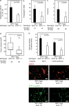
Blunted ALK1 signaling retards the angiogenic switch and tumor growth in the RIP1-Tag2 model. (a) Quantitative RT-PCR analysis of the expression of ALK1 in tumors from RIP1-Tag2; Alk1+/− mice compared with that of WT littermates (analysis independently performed at least three times). (b–d) Quantification of the number of angiogenic islets (b), the number of tumors (c), and total tumor burden (d) in RIP1-Tag2; Alk1wt mice and RIP1-Tag2; Alk1+/− mice. Box boundaries represent the interquartile range and the bars represent the full range. Solid and dotted line represents median and mean tumor volume, respectively. Circles denote statistical outliers (mean ± 2 SD). (e) Quantification of the vascular density, total (by CD31 immunostaining), and vessels perfused (by FITC-conjugated lectin), as a percentage of the lesional area, both in RIP1-Tag2; Alk1wt mice and RIP1-Tag2; Alk1+/− mice (n = 5 mice for each analysis; CD31, red; FITC-lectin, green). Error bars show the mean ± SD. Bars, 50 µm.
Figure 3.
Figure 3.
RAP-041 neutralizes BMP9 activity and inhibits angiogenic sprouting in vitro and in vivo. (a–c) Assessment of the capacity of RAP-041 to neutralize BMP9 (a), BMP10 (b), and TGF-β (c) by transcription activation luciferase reporter assays in C2C12 cells. Normalized promoter activity is plotted in arbitrary units as mean values from triplicate determinations (analyses independently performed at least three times). (d) Immunoblot analysis of the neutralizing activity of RAP-041 on BMP9-induced phosphorylation of Smad1 (pSmad1) in HUVEC (top) and on TGF-β–induced phosphorylation of Smad2 (pSmad2) and Smad3 (pSmad3) in C2C12 cells (bottom; analyses independently performed at least three times). The antibody used to detect pSmad3 (bottom band) also reacts with pSmad1 (top band). Immunoblotting for β-actin was used to ensure equal loading. Black lines indicate that intervening lanes have been spliced out. (e–g) Assessment of inhibition by RAP-041 of HUVEC tube formation (e) and spheroid sprouting (f; analyses independently performed at least three times) and VEGF- and FGF-induced vessels grown in subcutaneous Matrigel plugs (g; n = 5 mice per group). Error bars show the mean ± SD.
Figure 4.
Figure 4.
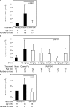
Pharmacological inhibition of ALK1 signaling retards tumor growth in the RIP1-Tag2 model. (a) Quantification of the total tumor burden of RIP1-Tag2 mice at 10 wk of age or after a 2-wk treatment with 12 mg/kg control Fc protein or RAP-041. (b) RAP-041 dose-response curve upon intraperitoneal injection of 1–24 mg/kg for 2 wk (10–12 wk of age). To allow a direct comparison, data for the cohort aged 10 wk, the control Fc cohort, and the 12-mg/kg cohort have been duplicated from a. (c) Quantification of the total tumor burden of RIP1-Tag2 mice at 12 wk of age or after a 2-wk treatment with 12 mg/kg of control Fc protein or RAP-041. Error bars show the mean ± SD.
Figure 5.
Figure 5.
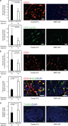
Treatment with RAP-041 impairs tumor angiogenesis in vivo. (a and b) Quantification of the vascular density by CD31 immunostaining (a) and by FITC-lectin perfusion (b) as a percentage of the lesional area, both in tumors from control Fc or RAP-041–treated mice of indicated age. (c) Quantification of pericyte coverage (number of NG2+ cells divided by number of CD31+ cells) of the vessels, as a percentage of the lesional area, of tumors from control Fc or RAP-041–treated mice. (d) Apoptotic index of tumors from control Fc or RAP-041–treated mice. n = 5 mice for each analysis; 10–15 high-power fields were scored for each mouse. Error bars show the mean ± SD. Bars, 50 µm.
Figure 6.
Figure 6.
BMP9 and TGF-β synergistically regulate endothelial cell function. (a) Quantification of the proliferative index of endothelial cells (presented as a mean for the three endothelial cell lines MS1, bEND.3, and TIME) as a percentage of Ki67 positively stained cells over the total number of cells upon no treatment or addition of different treatments with TGF-β, BMP9, VEGF-A, and combinations thereof. (b) Immunostaining for the endothelial cell marker CD31 (brown) of subcutaneously injected Matrigel plugs containing VEGF-A + bFGF, TGF-β + BMP9, RAP-041, and combinations thereof. (c) Quantification of the vascular ingrowth, as calculated by CD31 immunostaining in subcutaneously injected Matrigel plugs containing the factors indicated. (d) Ex vivo co-culture of RIP1-Tag2 angiogenic islets and MS1 endothelial cells on Matrigel upon absence or presence of treatment with TGF-β, BMP9, RAP-041, and combinations thereof. All analyses were independently performed at least three times. (e) Quantification of the area covered by migrating/sprouting endothelial cells bordering the angiogenic islet. Each mean represents quantification of five to eight angiogenic islets per condition. Error bars show the mean ± SD. Bars, 50 µm.
Figure 7.
Figure 7.
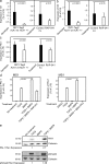
Both ALK1 and ALK5 target gene expression is down-regulated by blunted ALK1 signaling. (a–c) Id1 (a), Id3 (b), and PAI-1 (c) mRNA expression in tumors from RIP1-Tag2; Alk1+/− mice compared with that of WT littermates at 12 wk of age, as well as RIP1-Tag2 mice treated with control Fc (Ctrl Fc) protein or RAP-041 between 10 and 12 wk of age. The values for each gene represent mean and SD of at least three independent experiments with three to seven tumors per experimental condition. (d) Expression of PAI-1 mRNA in MS1 endothelial cells treated with TGF-β, BMP9, RAP-041, SB431542, and combinations thereof. Error bars show the mean ± SD. (e) Western blot analysis of PAI-1 protein levels and phosphorylated Smad2 (pSmad2) levels in lysates from MS1 cells subjected to single or combined stimulation of TGF-β and BMP9. Relative expression levels were calculated by densitometric quantification of PAI-1 or pSmad2 relative to the reference protein calnexin. All analyses were independently performed at least three times.

Similar articles

Cited by

References

    1. Arbiser J.L., Moses M.A., Fernandez C.A., Ghiso N., Cao Y., Klauber N., Frank D., Brownlee M., Flynn E., Parangi S., et al. 1997. Oncogenic H-ras stimulates tumor angiogenesis by two distinct pathways. Proc. Natl. Acad. Sci. USA. 94:861–866 10.1073/pnas.94.3.861 - DOI - PMC - PubMed
    1. Arthur H.M., Ure J., Smith A.J., Renforth G., Wilson D.I., Torsney E., Charlton R., Parums D.V., Jowett T., Marchuk D.A., et al. 2000. Endoglin, an ancillary TGFβ receptor, is required for extraembryonic angiogenesis and plays a key role in heart development. Dev. Biol. 217:42–53 10.1006/dbio.1999.9534 - DOI - PubMed
    1. Bajou K., Peng H., Laug W.E., Maillard C., Noel A., Foidart J.M., Martial J.A., DeClerck Y.A. 2008. Plasminogen activator inhibitor-1 protects endothelial cells from FasL-mediated apoptosis. Cancer Cell. 14:324–334 10.1016/j.ccr.2008.08.012 - DOI - PMC - PubMed
    1. Bergers G., Hanahan D. 2008. Modes of resistance to anti-angiogenic therapy. Nat. Rev. Cancer. 8:592–603 10.1038/nrc2442 - DOI - PMC - PubMed
    1. Bergers G., Javaherian K., Lo K.M., Folkman J., Hanahan D. 1999. Effects of angiogenesis inhibitors on multistage carcinogenesis in mice. Science. 284:808–812 10.1126/science.284.5415.808 - DOI - PubMed

Publication types

MeSH terms

Substances