. 2010 Jan 11;188(1):115-30.
doi: 10.1083/jcb.200903137.

Neuropilin-2 mediates VEGF-C-induced lymphatic sprouting together with VEGFR3

Affiliations

Neuropilin-2 mediates VEGF-C-induced lymphatic sprouting together with VEGFR3

Yunling Xu et al. J Cell Biol. .

Abstract

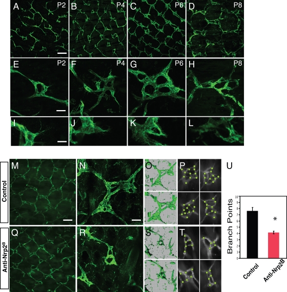
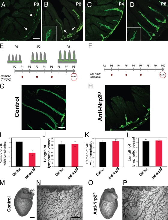
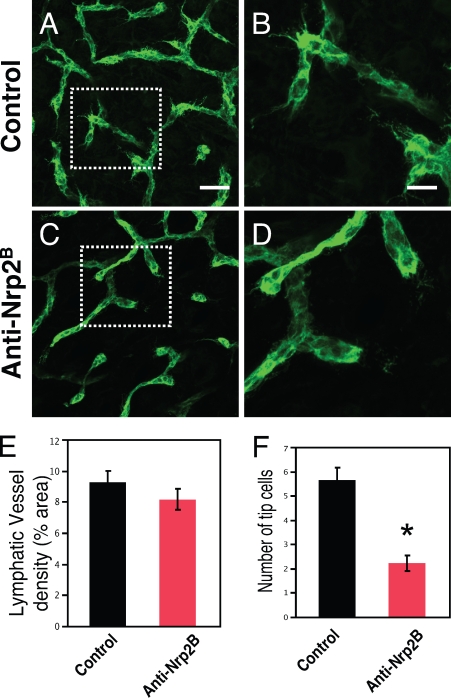
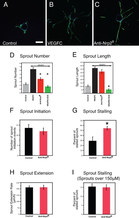
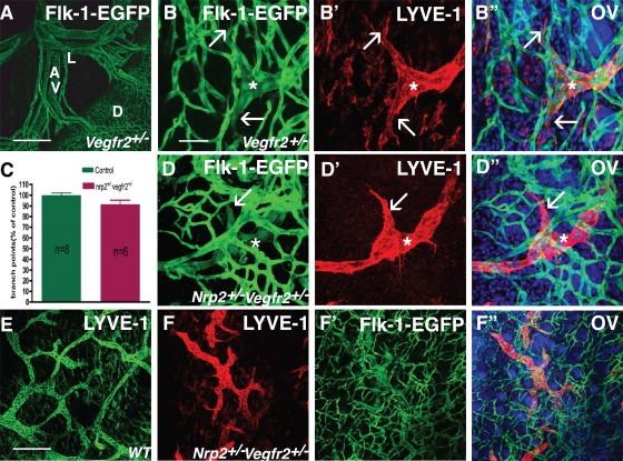
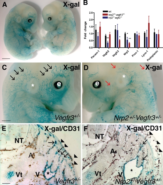
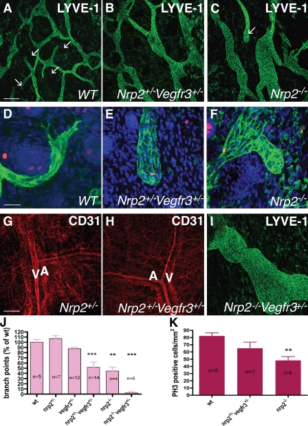
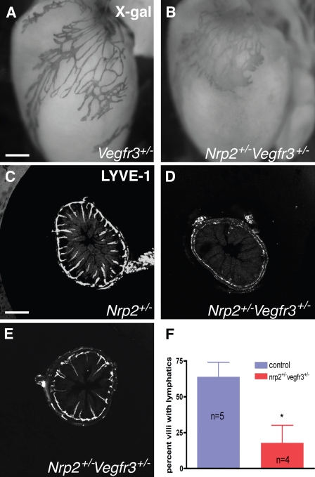
Vascular sprouting is a key process-driving development of the vascular system. In this study, we show that neuropilin-2 (Nrp2), a transmembrane receptor for the lymphangiogenic vascular endothelial growth factor C (VEGF-C), plays an important role in lymphatic vessel sprouting. Blocking VEGF-C binding to Nrp2 using antibodies specifically inhibits sprouting of developing lymphatic endothelial tip cells in vivo. In vitro analyses show that Nrp2 modulates lymphatic endothelial tip cell extension and prevents tip cell stalling and retraction during vascular sprout formation. Genetic deletion of Nrp2 reproduces the sprouting defects seen after antibody treatment. To investigate whether this defect depends on Nrp2 interaction with VEGF receptor 2 (VEGFR2) and/or 3, we intercrossed heterozygous mice lacking one allele of these receptors. Double-heterozygous nrp2vegfr2 mice develop normally without detectable lymphatic sprouting defects. In contrast, double-heterozygote nrp2vegfr3 mice show a reduction of lymphatic vessel sprouting and decreased lymph vessel branching in adult organs. Thus, interaction between Nrp2 and VEGFR3 mediates proper lymphatic vessel sprouting in response to VEGF-C.


Figures

Figure 1.
Figure 1.
Nrp2 inhibition results in abnormal tail dermal lymphangiogenesis. (A–L) Developmental time course of tail dermal lymphangiogenesis by whole-mount LYVE-1 immunohistochemistry at P2, 4, 6, and 8. The tails are imaged to show the overall lymphatic network pattern (A–D), to demonstrate representative ring complexes (E–H), and to demonstrate tip cells extending from the complexes (I–L). (M–U) Treatment with anti-Nrp2B results in alteration of lymphangiogenesis. The lymphatics in anti-Nrp2B–treated tails at P8 after treatment at P1, 3, and 5. 3D projections of the confocal images in control (O) and anti-Nrp2B–treated ring complexes (S) in N and R, respectively. The bottom projection is rotated 45° to provide depth perspective. (P and T) Representative examples of junctions with dots to denote tallied branch points in control (P) and anti-Nrp2B (T)–treated tails. (U) Quantification of branch points in ring complexes of control and anti-Nrp2B–treated tails (n = 10 junctions/animal for six animals per condition; *, P < 0.0001). Error bars indicate SEM. Bars: (A–D, M, and Q) 250 µm; (E–H) 60 µm; (I–L) 25 µm; (N–R) 75 µm.
Figure 2.
Figure 2.
Nrp2 inhibition reduces lymphatic sprouting in developing intestinal and epicardial lymphatics. (A–D) Developmental time course of intestinal lymphangiogenesis by LYVE-1 immunohistochemistry. (A) At P0, lymphatics are restricted to the submucosa (arrows). (B) By P2, lymphatic branches have extended into a portion of some villi, and newly formed sprouts (arrows) can be seen to form on the submucosal lymphatic vessel adjacent to other villi. Tip cells with filopodia (inset) are present on the growing lacteals. (C and D) By P4 (C), most villi have a developing lacteal, which extends to the villus tip by P8 (D). Tip cells (inset) can still be observed at the ends of the vessels. (E) Scheme of the time course of lymphatic sprouting into intestinal villi and representation of experiments shown in G, I, and J. Anti-Nrp2B was injected i.p. at P1, 3, and 5 (red dots), animals were sacrificed at P8, and tissues were analyzed. (F) Schematic representation of experiment shown in H, K, and L. Anti-Nrp2B was injected i.p. at P3, 5, and 7 (red dots), animals were sacrificed at P10, and tissues were analyzed. (G–J) Analysis of intestines from the experiment depicted in E. (G and H) Control-treated intestines (G) have a normal-appearing lymphatic pattern in contrast to anti-Nrp2B–treated intestines (H) in which a larger portion of villi lack lacteals. (I and J) Quantification of the percent of villi that have lacteals and villi length. (I–L) 50 villi per animal for six animals per treatment condition were analyzed. (M–P) Control (M and N) and anti-Nrp2B (O and P)–treated hearts showing reduced branching and increased vessel thickness in portions of the anti-Nrp2B–treated hearts. Error bars indicate SEM. Bars: (A–D) 250 µm; (G and H) 125 µm; (M–P) 400 µm.
Figure 3.
Figure 3.
Anti-Nrp2B treatment reduces tip cell number in dermal lymphatics. (A–D) Control (A and B) and anti-Nrp2B (C and D)–treated skin showing a reduction in the number of tip cells. The boxed areas in A and C are shown at higher magnifications in B and D, respectively. (E) No change in lymphatic vessel density was noted between control and anti-Nrp2B–treated samples. (F) A significant reduction in the number of tip cells per high powered field was noted between control and anti-Nrp2B–treated samples. Error bars indicate SEM. *, P < 0.01. Bars: (A and C) 150 µm; (B and D) 70 µm.
Figure 4.
Figure 4.
Anti-Nrp2B treatment in vitro results in reduced sprout formation and altered tip cell behavior. (A–C) Representative examples of LECs sprouting from coated beads in control (A), VEGF-C (B)–, or anti-Nrp2B (C)–treated cultures stained with anti–LYVE-1. (D) Sprout number is significantly reduced with anti-Nrp2B or VEGFR3 ECD treatment compared with VEGF-C treatment alone. (E) Sprout length is not reduced with anti-Nrp2B treatment but is reduced by VEGFR3 ECD treatment compared with VEGF-C treatment alone (n = 5 beads/well for 10 wells). (F–I) Quantification of sprout initiation events (F), sprout-stalling events (G), and sprout extension rates from live imaging experiments. Error bars indicate SEM. *, P < 0.01. Bar, 150 µm.
Figure 5.
Figure 5.
Normal lymphatic development in double-heterozygous nrp2+/−vegfr2+/− mice. (A) EGFP staining of mesenteric vessels in a heterozygous vegfr2-egfp mouse pup at P0. Note green fluorescence in capillaries covering the surface of the duodenum and in mesenteric arteries, veins, and lymphatic vessels. D, duodenum; A, mesenteric arteries; V, veins; L, lymphatic vessels. (B–B″) EGFP–LYVE-1 double staining of tail skin at P1. Note Flk-1–EGFP expression in a lymphatic vessel sprout (asterisks) and in filopodia-extending tips (arrows). (C) Quantification of lymph vessel branch points. Error bars indicate SEM. (D–D″) Normal appearance of lymphatic vessel sprouts (asterisks) and filopodia-extending tips (arrows) in P1 tail skin in double-heterozygous nrp2+/−vegfr2+/− mice. (E and F) Lower magnification views of LYVE-1–positive lymphatic vessels in wild-type (E) and nrp2+/−vegfr2+/− dorsal trunk skin. WT, wild type. (F’–F″) Flk-1/VEGFR2-EGFP–LYVE-1 double labeling. OV, overlay. Bars: (A) 200 µm; (B–D″) 50 µm; (E–F″) 200 µm.
Figure 6.
Figure 6.
Abnormal lymphatic development in double-heterozygous nrp2+/−vegfr3+/− mice. (A) X-gal staining (blue) of E13.5 vegfr3+/− (left) and double-heterozygous nrp2+/−vegfr3+/− (right) littermate embryos. (B) expression levels of lymphatic marker genes as measured by quantitative PCR in RNA isolated from hearts of wild-type and double-heterozygote nrp2+/−vegfr2+/− and nrp2+/−vegfr3+/− mice. Values significantly different from wild-type mice (*, P > 0.05; **, P > 0.001) by Student’s t test are shown. Error bars indicate SEM. (C and D) Higher magnification of heads of embryos shown in A. Note numerous lymphatic vessel sprouts in vegfr3+/− (black arrows) and fewer enlarged lymph vessel sprouts in nrp2+/−vegfr3+/− (red arrows). e, eye; *, ear. (E and F) Transverse section through the neck of E13.5 embryos double stained with X-gal (blue) and CD-31 (brown). Note similar development of CD-31–positive arteries, veins, and skin capillaries (arrowheads) in vegfr3+/− (E) and nrp2+/−vegfr3+/− (F). Note enlarged jugular lymph sacs (asterisks) in nrp2+/−vegfr3+/− (F) compared with vegfr3+/− (E). Lymph vessels sprouting from the lymph sac toward the skin are less numerous in nrp2+/−vegfr3+/− compared with vegfr3+/− (arrows). A, arteries; V, veins; NT, neural tube; Vt, vertebra. Bars: (A) 1.4 mm; (C and D) 0.8 mm; (E and F) 150 µm.
Figure 7.
Figure 7.
Defective lymphatic sprouting in nrp2+/−vegfr3+/− mice. (A–C) Confocal images of LYVE-1–stained lymph vasculature in the dorsal trunk skin at P0. Note enlarged, poorly branched vessels in nrp2+/−vegfr3+/− (B) and nrp2−/− (C) compared with wild type (A). Several lymphatic tips are present in wild type (A, arrows), none are present in nrp2+/−vegfr3+/− (B), and one is present in nrp2−/− (C, arrow). (D–F) LYVE-1 (green)/phospho–histone H3 (red) DAPI (blue) staining of P0 skin; high magnification views of lymphatic tips are shown. Note filopodial-extending tips composed of several cells in wild type (D), whereas both nrp2+/−vegfr3+/− (E) and nrp2−/− (F) show blunt-ended, enlarged tips devoid of filopodial extensions. (G and H) CD31 staining shows similar patterning of blood vasculature in the skin of mice with the indicated genotypes. A, arteries; V, veins. (I) Dorsal trunk skin at P0 in a nrp2−/−vegfr3+/− mouse. Note enlarged lymphatic vessel. (J) Quantification of lymphatic vessel branch points in mice of the indicated genotypes. (K) Replication of LECs. The number of phospho–histone H3 (PH3)-positive nuclei per millimeter squared of LYVE-1–positive vessels was counted. WT, wild type. Error bars indicate SEM. **, P < 0.01; ***, P < 0.001. Bars: (A–C) 200 µm; (D–F) 50 µm; (G–I) 200 µm.
Figure 8.
Figure 8.
Tip cell abnormalities in nrp2+/−vegfr3+/− mice are similar to those obtained after anti-Nrp2B treatment. (A and B) X-gal staining of P5 hearts from mice of the indicated genotypes. Note regular branching of lymphatics in the vegfr3+/− heart (A) but enlarged, poorly branched vessels devoid of sprouting tips in the double-heterozygote heart (B). (C–F) Lymphatic sprouting defects in the small intestine at P4. LYVE-1–stained sections show presence of lymphatics in almost all villi of wild-type mice (C) but virtually no sprouts in most sections of the double-heterozygous mice (D). Only some sections show a portion of the villi-containing lymphatics (E), which are of normal length compared with wild type (C). (F) Quantification of the percentage of villi-containing lymphatics. *, P < 0.01. Error bars indicate SEM. Bars: (A and B) 210 µm; (C–E) 200 µm.

Similar articles

Cited by

References

    1. Affolter M., Caussinus E. 2008. Tracheal branching morphogenesis in Drosophila: new insights into cell behaviour and organ architecture. Development. 135:2055–2064 10.1242/dev.014498 - DOI - PubMed
    1. Alitalo K., Tammela T., Petrova T.V. 2005. Lymphangiogenesis in development and human disease. Nature. 438:946–953 10.1038/nature04480 - DOI - PubMed
    1. Bagri A., Cheng H.J., Yaron A., Pleasure S.J., Tessier-Lavigne M. 2003. Stereotyped pruning of long hippocampal axon branches triggered by retraction inducers of the semaphorin family. Cell. 113:285–299 10.1016/S0092-8674(03)00267-8 - DOI - PubMed
    1. Baluk P., Fuxe J., Hashizume H., Romano T., Lashnits E., Butz S., Vestweber D., Corada M., Molendini C., Dejana E., McDonald D.M. 2007. Functionally specialized junctions between endothelial cells of lymphatic vessels. J. Exp. Med. 204:2349–2362 10.1084/jem.20062596 - DOI - PMC - PubMed
    1. Cai H., Reed R.R. 1999. Cloning and characterization of neuropilin-1-interacting protein: a PSD-95/Dlg/ZO-1 domain-containing protein that interacts with the cytoplasmic domain of neuropilin-1. J. Neurosci. 19:6519–6527 - PMC - PubMed

Publication types

MeSH terms

Substances