Comparative Study
. 2010 Feb 15;184(4):1821-8.
doi: 10.4049/jimmunol.0902869. Epub 2010 Jan 11.

Dynamic equilibrium of B7-1 dimers and monomers differentially affects immunological synapse formation and T cell activation in response to TCR/CD28 stimulation

Affiliations
Comparative Study

Dynamic equilibrium of B7-1 dimers and monomers differentially affects immunological synapse formation and T cell activation in response to TCR/CD28 stimulation

Sumeena Bhatia et al. J Immunol. .

Abstract

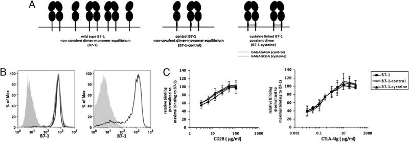
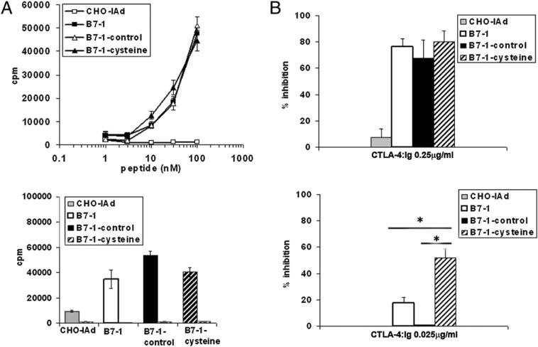
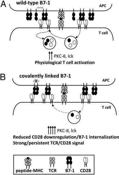
Under steady-state conditions, B7-1 is present as a mixed population of noncovalent dimers and monomers on the cell surface. In this study, we examined the physiological significance of this unique dimer-monomer equilibrium state of B7-1. We demonstrate that altering B7-1 to create a uniformly covalent dimeric state results in enhanced CD28-mediated formation of T cell-APC conjugates. The enhanced T cell-APC conjugate formation correlates with persistent concentration of signaling molecules PKC- and lck at the immunological synapse. In contrast, T cell acquisition of B7-1 from APCs, an event that occurs as a consequence of CD28 engagement with B7-1/B7-2 and is thought to play a role in the dissociation of T cell-APC conjugates, is highly reduced when B7-1 is present in the covalently dimeric state. The ability of covalently dimeric and wild type B7-1 to costimulate Ag-specific T cell proliferation was also assessed. In contrast to the enhanced ability of dimeric B7-1 to support conjugate formation and early parameters of T cell signaling, sensitivity to competitive inhibition by soluble CTLA-4-Ig indicated that the covalent dimeric form of B7-1 is less efficient in costimulating T cell proliferation. These findings suggest a novel model in which optimal T cell costimulatory function of B7-1 requires high-avidity CD28 engagement by dimeric B7-1, followed by dissociation of these noncovalent B7-1 dimers, facilitating downregulation of CD28 and internalization of B7-1. These events regulate signaling through TCR/CD28 to maximize T cell activation to proliferation.


Figures

FIGURE 1
FIGURE 1
Characterization of WT B7-1, B7-1–control, and B7-1–cysteine. A, Schematic representation of WT B7-1 or its mutants, engineered with an 8-aa residue linker sequence in the extracellular stalk region, with (B7-1–cysteine) or without (B7-1–control) cysteine. B, Left panel: Graphs showing the expression of WT B7-1 (black), B7-1–control (light gray), and B7-1–cysteine (dark gray) on CHO-IAd cells. Shaded curve represents staining for B7-1 on untransfected CHO-IAd cells. Right panel: Staining of B7-1 (black) on activated splenic dendritic cells; shaded curve represents the isotype control. C, Quantitative analysis of CD28Ig (left graph) and CTLA-4Ig (right graph) binding to WT B7-1, B7-1–control, and B7-1–cysteine expressing stable CHOIAd transfectants. The relative binding at each concentration of CD28Ig or CTLA-4Ig was calculated as [fluorescence intensity B7-1mutant/maximal fluorescence intensity B7-1WT] × 100. The results are the mean of four independent experiments; and the error bars represent the SE of the mean.
FIGURE 2
FIGURE 2
Expression of obligate covalently linked B7-1 dimers increases CD28-mediated adhesion to T cells. A, CHO-IAd cells expressing the covalently linked dimeric B7-1 show increased adhesion to T cells. CHO-IAd cells untransfected (top row) or expressing WT B7-1 (second row), B7-1–control (third row), or B7-1–cysteine (bottom row) were pulsed with a titrated concentration of OVA peptide, labeled with CMTMR, and mixed with CFSE-labeled DO11.10 TCR Tg T cells. The CMTMR+CFSE+ CHO-IAd–T cell conjugates were scored by flow cytometry. The numbers in the top right corner represent the percentage of T cells as T cell–APC conjugates. B, Quantification of CHO-IAd–T cell conjugates as a percentage of total CD4 T cells. The results and error bars represent the means and SD from four independent experiments. *p < 0.05.
FIGURE 3
FIGURE 3
Persistent concentration of PKC-θ and lck at the IS in the presence of covalently dimeric B7-1. A, Peptide-pulsed B7-1–transfected CHO-IAd cells were fixed after mixing with DO11.10 CD4 T cells. The cells were stained for intracellular lck (green) and PKC-θ (blue). Arrow indicates the line of measurement of fluorescence intensity for PKC-θ or lck from distal area to contact area for the T cell–APC conjugates. The graphs show the fluorescence intensities of PKC-θ or lck along the marked arrow, extending from a region on the distal T cell membrane to a region on the T cell membrane in contact with APC. Fluorescence intensity was calculated as the area under the curve representing the IS or the distal membrane. The ratio of fluorescence intensity for the IS/fluorescence intensities for the distal membrane was used to calculate fold enrichment of the signaling molecules at the IS. T cell–APC conjugates that showed >2-fold enrichment of PKC-θ in the IS were scored as positive events. A representative image of T cells with >2-fold enrichment of PKC-θ (20 min) or lck (7 min) at the IS is shown in the respective upper panels, whereas the lower panels represent T cells that did not enrich PKC-θ or lck in the IS. B, Quantification of T cell conjugates with concentration of PKC-θ or lck at the IS. Left graph: Percentage of T cells showing concentration of PKC-θ in the IS at 2 and 20 min in the presence of CHO-IAd (n = 167; 173), B7-1–control (n = 207; 153), or B7-1–cysteine (n = 190; 182). The results for PKC-θ represent the means and SE from four independent experiments. Right graph: Percentage of T cells showing >1.5-fold accumulation of lck in the IS at 2 and 7 min in the presence of CHO-IAd (n = 97; 81), B7-1–control (n = 123; 138), or B7-1–cysteine (n = 111; 147). The results for lck represent the means and SE from three independent experiments. C, Upper left: Representative image of a mature IS characterized by PKC-θ and LFA-1 segregation in the IS of DO11.10.DKO T cells. Bottom left: Pseudo-colored 2.5D plot showing segregation of PKC-θ and LFA-1 in the IS. Right: Three-dimensional reconstruction of z-stacks of images showing the central localization of PKC-θ surrounded by peripheral LFA-1, characteristic of a mature IS. D, Quantification of T cell– APC conjugates that showed segregation of PKC-θ and LFA-1 in the IS. Thirty to 40 conjugates were visualized per group per experiment. The results are the mean and SE of three (for DO11.10.DKO) or two (DO11.10.CD28KO) independent experiments. *p < 0.05. ns, not significant.
FIGURE 4
FIGURE 4
Reduced acquisition of covalently dimeric B7-1 by T cells. A, Peptide-pulsed CHO-IAd–B7-1 (left panels) or CHO-IAd–B7-1-cysteine (right panels) cells were fixed at 30 min (first and second rows) or 3 h (third and fourth rows) after mixing with DO11.10 CD4 T (first and third rows) or DO11.10. CD28−/− CD4 T (second and fourth rows) cells. The cells were stained for intracellular B7-1 (green) and CD4 (red). T cells in T cell–APC conjugates that showed B7-1 fluorescence (intracellular or cell surface associated) greater than the threshold value obtained from T cells only were scored positive for the acquisition of B7-1. B, Left panel: Quantification of T cells in conjugates that show acquisition of B7-1 3 h after T cell–APC conjugate formation in the absence or presence of OVA peptide. The results are mean and SD from six independent experiments. Right panel: Quantification of total B7-1 fluorescence per T cell. Each circle represents one T cell. Forty to 50 conjugates were visualized in each group. The results are representative of four independent experiments. **p , 0.01. C, Quantification of T cells in conjugates that show acquisition of B7-1 at 30 min (left graph) or 3 h (right graph) after T cell–APC conjugate formation. The black bars indicate WT DO11.10 TCRTg CD4 T cells, whereas the red bars indicate DO11.10 TCR Tg CD28-deficient CD4 T cells. Forty to 50 conjugates were visualized in each group per experiment and is mean of two independent experiments; the circles represent individual experiments.
FIGURE 5
FIGURE 5
B7-1–cysteine is less efficient in costimulating T cell proliferation. A, Top panel: DO11.10 CD4+ T cells were stimulated with titrated concentrations of OVA peptide in the presence of CHO-IAd cells that were untransfected or expressing WT B7-1, B7-1–control, or B7-1–cysteine as APCs for 48 h, after which the cultures were pulsed with 1 μCi of [3H]thymidine for an additional 16 h. Bottom panel: CD4+ T cells from DO11.10 B7 DKO (black bars) or CD28 KO (gray hatched bars) mice were stimulated with 30 nM OVA peptide in the presence of CHO-IAd cells that were untransfected or expressing WT B7-1, B7-1–control, or B7-1–cysteine as APCs for 48 h and processed as described above. B, DO11.10 CD4+ T cells were stimulated with OVA peptide (30 nM) in the presence of CHO-IAd cells that were untransfected or expressing WT B7-1, B7-1–control, or B7-1–cysteine as APCs in the presence of 0.25 μg/ml (top panel) or 0.025 μg/ml (bottom graph) of CTLA-4–Ig for 48h, after which the cultures were pulsed with 1 μCi of [3H]thymidine for an additional 16 h. Triplicate wells were harvested and counted. The percentage inhibition was calculated as {[(CPM without CTLA-4Ig) – (CPM with CTLA-4Ig)]/(CPM with CTLA-4Ig)} × 100. The results are the mean and SE of four independent experiments. *p < 0.05.
FIGURE 6
FIGURE 6
Model for the interaction of B7-1 with CD28. A, Upon binding of noncovalent dimers of B7-1 to CD28, a fraction of CD28 is internalized, part of which is recycled back to the cell surface, whereas the remaining is targeted to lysosomes for degradation. Thus, the cell surface expression of CD28 is regulated by multiple mechanisms. The internalization of CD28 is also concomitant with the absorption of B7-1. Results of the present studies comparing noncovalent B7-1 dimers (B7-1–control) and covalently linked B7-1 dimers (B7-1–cysteine) suggest that this process of internalization of CD28 and acquisition of B7-1 may involve a key step of dissociation of noncovalent dimers of B7-1 (double-sided arrow). B, In the presence of a covalently linked dimeric form of B7-1, there is reduced acquisition of B7-1 and, presumably, reduced internalization of CD28. In the absence of dissociation of B7-1 dimers, signaling events, including IS localization of PKC-θ and lck, are increased and prolonged as the result of enhanced and/or sustained signaling via TCR/CD28.

Similar articles

Cited by

References

    1. Greenwald RJ, Freeman GJ, Sharpe AH. The B7 family revisited. Annu. Rev. Immunol. 2005;23:515–548. - PubMed
    1. Keir ME, Sharpe AH. The B7/CD28 costimulatory family in autoimmunity. Immunol. Rev. 2005;204:128–143. - PubMed
    1. Hathcock KS, Laszlo G, Pucillo C, Linsley P, Hodes RJ. Comparative analysis of B7-1 and B7-2 costimulatory ligands: expression and function. J. Exp. Med. 1994;180:631–640. - PMC - PubMed
    1. Inaba K, Witmer-Pack M, Inaba M, Hathcock KS, Sakuta H, Azuma M, Yagita H, Okumura K, Linsley PS, Ikehara S, et al. The tissue distribution of the B7-2 costimulator in mice: abundant expression on dendritic cells in situ and during maturation in vitro. J. Exp. Med. 1994;180:1849–1860. - PMC - PubMed
    1. Collins AV, Brodie DW, Gilbert RJ, Iaboni A, Manso-Sancho R, Walse B, Stuart DI, van der Merwe PA, Davis SJ. The interaction properties of costimulatory molecules revisited. Immunity. 2002;17:201–210. - PubMed

Publication types

MeSH terms