

Donepezil treatment and changes in hippocampal structure in very mild Alzheimer disease
- PMID: 20065136
- PMCID: PMC2855123
- DOI: 10.1001/archneurol.2009.292
Donepezil treatment and changes in hippocampal structure in very mild Alzheimer disease
Abstract
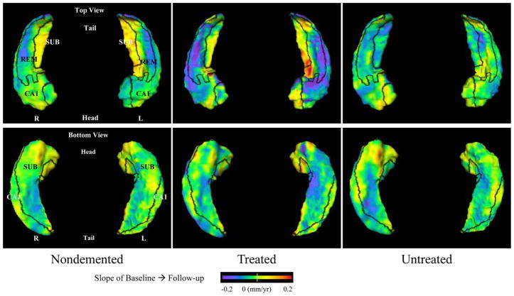
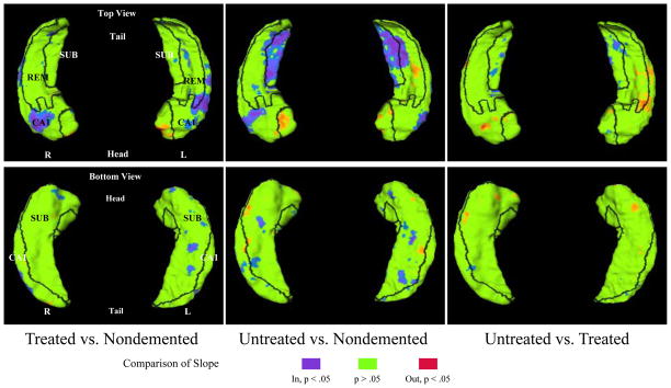
Objective: To compare longitudinal changes in the hippocampal structure in subjects with very mild dementia of the Alzheimer type (DAT) treated with donepezil hydrochloride, untreated subjects with very mild DAT, and controls without dementia.
Design: MPRAGE sequences were collected approximately 2 years apart on two 1.5-T magnetic resonance imaging systems, yielding 2 cohorts. Large-deformation high-dimensional brain mapping was used to compute deformation of hippocampal subfields.
Setting: A dementia clinic at Washington University School of Medicine.
Patients or other participants: Subjects came from 2 sources: 18 untreated subjects with DAT and 26 controls were drawn from a previous longitudinal study; 18 treated subjects with DAT were studied prospectively, and 44 controls were drawn from a longitudinal study from the same period. Intervention Patients were prescribed donepezil by their physician.
Main outcome measures: Hippocampal volume loss and surface deformation.
Results: There was no significant cohort effect at baseline; therefore, the 2 groups of control subjects were combined. The potential confounding effect of cohort/scanner was dealt with by including it as a covariate in statistical tests. There was no significant group effect in the rate of change of hippocampal volume or subfield deformation. Further exploration showed that compared with the untreated subjects with DAT, the treated subjects with DAT did not differ in the rate of change in any of the hippocampal measures. They also did not differ from the controls, while the untreated subjects with DAT differed from the controls in the rates of change of hippocampal volume and CA1 and subiculum subfield deformations.
Conclusions: Treatment with donepezil did not alter the progression of hippocampal deformation in subjects with DAT in this study. Small sample size may have contributed to this outcome.
Figures
Similar articles
-
Does donepezil treatment slow the progression of hippocampal atrophy in patients with Alzheimer's disease?Am J Psychiatry. 2005 Apr;162(4):676-82. doi: 10.1176/appi.ajp.162.4.676. Am J Psychiatry. 2005. PMID: 15800138 Clinical Trial.
-
Neuroanatomical predictors of response to donepezil therapy in patients with dementia.Arch Neurol. 2005 Nov;62(11):1718-22. doi: 10.1001/archneur.62.11.1718. Arch Neurol. 2005. PMID: 16286546 Clinical Trial.
-
Donepezil effects on hippocampal and prefrontal functional connectivity in Alzheimer's disease: preliminary report.J Alzheimers Dis. 2012;31 Suppl 3(0 3):S221-6. doi: 10.3233/JAD-2012-120709. J Alzheimers Dis. 2012. PMID: 22886013 Free PMC article.
-
Donepezil: an update.Expert Opin Pharmacother. 2007 May;8(7):1011-23. doi: 10.1517/14656566.8.7.1011. Expert Opin Pharmacother. 2007. PMID: 17472546 Review.
-
[Nitrergic cerebrovascular regulation as affected by donepezil].Nihon Yakurigaku Zasshi. 2013 Mar;141(3):150-4. doi: 10.1254/fpj.141.150. Nihon Yakurigaku Zasshi. 2013. PMID: 23470480 Review. Japanese. No abstract available.
Cited by
-
Donepezil impairs memory in healthy older subjects: behavioural, EEG and simultaneous EEG/fMRI biomarkers.PLoS One. 2011;6(9):e24126. doi: 10.1371/journal.pone.0024126. Epub 2011 Sep 8. PLoS One. 2011. PMID: 21931653 Free PMC article. Clinical Trial.
-
A Systematic Review of Longitudinal Studies Which Measure Alzheimer's Disease Biomarkers.J Alzheimers Dis. 2017;59(4):1359-1379. doi: 10.3233/JAD-170261. J Alzheimers Dis. 2017. PMID: 28759968 Free PMC article.
-
Cholinesterase inhibitors for mild cognitive impairment.Cochrane Database Syst Rev. 2012 Sep 12;2012(9):CD009132. doi: 10.1002/14651858.CD009132.pub2. Cochrane Database Syst Rev. 2012. PMID: 22972133 Free PMC article.
-
Use of biomarkers in clinical trials of Alzheimer disease: from concept to application.Mol Diagn Ther. 2011 Dec 1;15(6):313-25. doi: 10.1007/BF03256467. Mol Diagn Ther. 2011. PMID: 22188635
-
A pilot study of brain morphometry following donepezil treatment in mild cognitive impairment: volume changes of cortical/subcortical regions and hippocampal subfields.Sci Rep. 2020 Jul 2;10(1):10912. doi: 10.1038/s41598-020-67873-y. Sci Rep. 2020. PMID: 32616841 Free PMC article.
References
-
- Cummings JL. Alzheimer’s disease. N Engl J Med. 2004 Jul 1;351(1):56–67. - PubMed
-
- Raina P, Santaguida P, Ismaila A, et al. Effectiveness of cholinesterase inhibitors and memantine for treating dementia: evidence review for a clinical practice guideline. Ann Intern Med. 2008 Mar 4;148(5):379–397. - PubMed
-
- Farlow MR, Lahiri DK, Poirier J, Davignon J, Schneider L, Hui SL. Treatment outcome of tacrine therapy depends on apolipoprotein genotype and gender of the subjects with Alzheimer’s disease. Neurology Mar. 1998;50(3):669–677. - PubMed
Publication types
MeSH terms
Substances
Grants and funding
LinkOut - more resources
Full Text Sources
Medical
Miscellaneous
