Potential aggregation prone regions in biotherapeutics: A survey of commercial monoclonal antibodies
- PMID: 20065649
- PMCID: PMC2726584
- DOI: 10.4161/mabs.1.3.8035
Potential aggregation prone regions in biotherapeutics: A survey of commercial monoclonal antibodies
Abstract
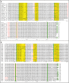
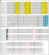
Aggregation of a biotherapeutic is of significant concern and judicious process and formulation development is required to minimize aggregate levels in the final product. Aggregation of a protein in solution is driven by intrinsic and extrinsic factors. In this work we have focused on aggregation as an intrinsic property of the molecule. We have studied the sequences and Fab structures of commercial and non-commercial antibody sequences for their vulnerability towards aggregation by using sequence based computational tools to identify potential aggregation-prone motifs or regions. The mAbs in our dataset contain 2 to 8 aggregation-prone motifs per heavy and light chain pair. Some of these motifs are located in variable domains, primarily in CDRs. Most aggregation-prone motifs are rich in beta branched aliphatic and aromatic residues. Hydroxyl-containing Ser/Thr residues are also found in several aggregation-prone motifs while charged residues are rare. The motifs found in light chain CDR3 are glutamine (Q)/asparagine (N) rich. These motifs are similar to the reported aggregation promoting regions found in prion and amyloidogenic proteins that are also rich in Q/N, aliphatic and aromatic residues. The implication is that one possible mechanism for aggregation of mAbs may be through formation of cross-beta structures and fibrils. Mapping on the available Fab-receptor/antigen complex structures reveals that these motifs in CDRs might also contribute significantly towards receptor/antigen binding. Our analysis identifies the opportunity and tools for simultaneous optimization of the therapeutic protein sequence for potency and specificity while reducing vulnerability towards aggregation.
Figures


References
-
- Kohler G, Milstein C. Continuous cultures of fused cells secreting antibody of predefined specificity. Nature. 1975;256:495–497. - PubMed
-
- McCafferty J, Griffiths AD, Winter G, Chiswell DJ. Phage antibodies: filamentous phage displaying antibody variable domains. Nature. 1990;348:552–554. - PubMed
-
- Reichert JM, Rosensweig CJ, Faden LB, Dewitz MC. Monoclonal antibody successes in the clinic. Nat Biotechnol. 2005;23:1073–1078. - PubMed
-
- Branden C, Tooze J. Introduction to Protein Structure. Garland Publishing; 1998.
Publication types
MeSH terms
Substances
LinkOut - more resources
Full Text Sources
Other Literature Sources
