

Ex vivo analysis of irradiated fingernails: chemical yields and properties of radiation-induced and mechanically-induced radicals
- PMID: 20065698
- PMCID: PMC3684967
- DOI: 10.1097/HP.0b013e3181b0c045
Ex vivo analysis of irradiated fingernails: chemical yields and properties of radiation-induced and mechanically-induced radicals
Abstract
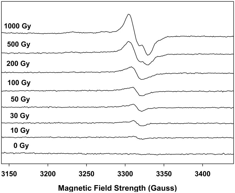
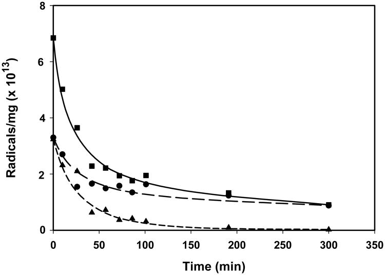
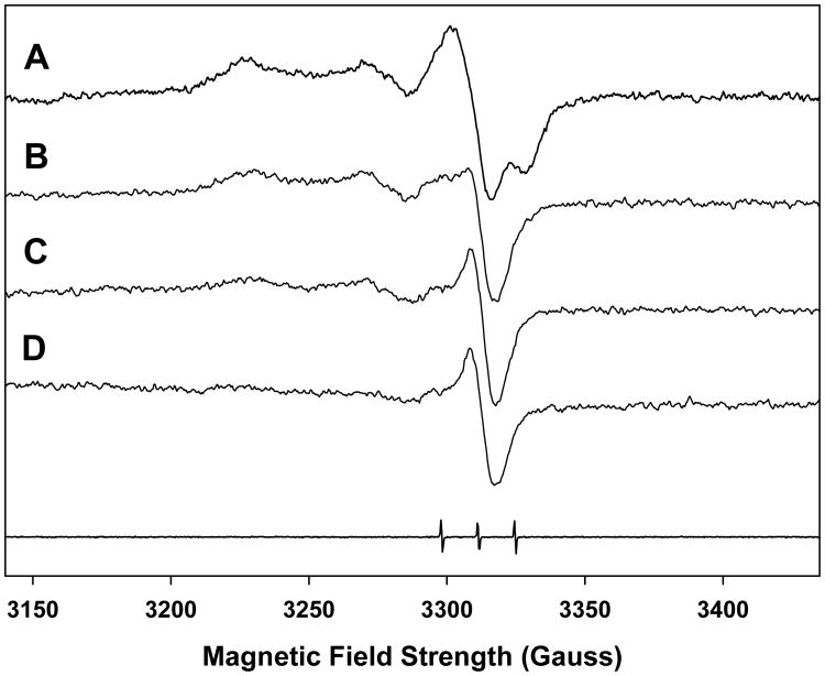
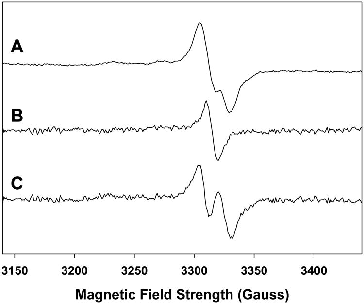
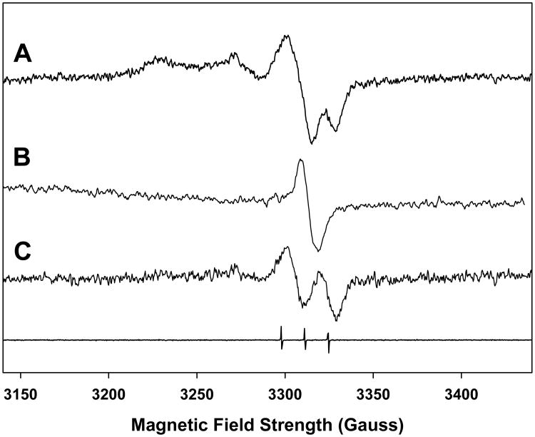
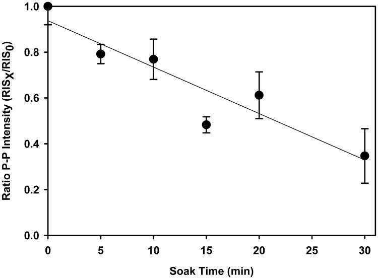
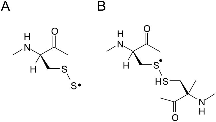
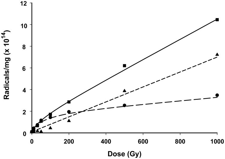
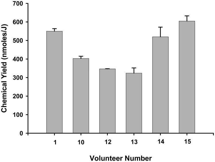
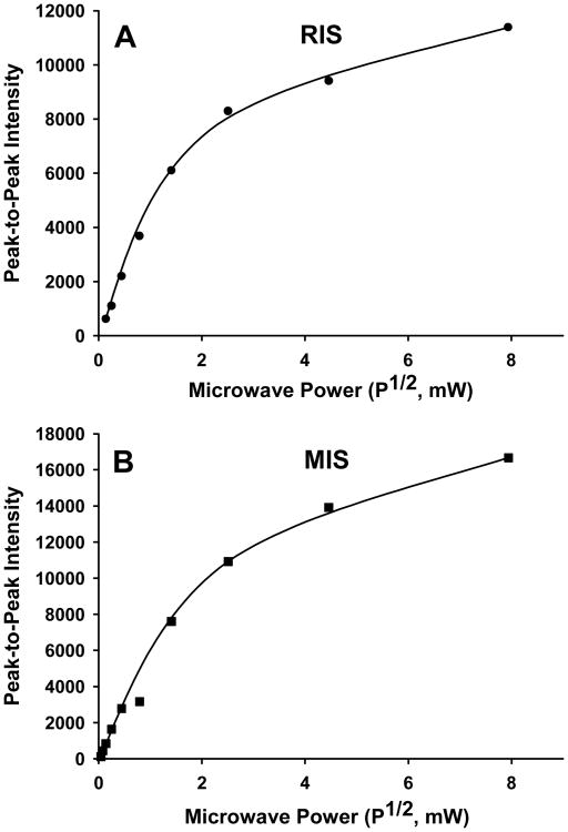
A qualitative and quantitative analysis of the radicals underlying the radiation-induced signal (RIS) in fingernails was conducted in an attempt to identify properties of these radicals that could be used for biodosimetry purposes. A qualitative analysis of RIS showed the presence of at least three components, two of which were observed at low doses (<50 Gy) and the third required higher doses (>500 Gy). The low dose signal, obtained by reconstruction, consists of a 10 gauss singlet at g = 2.0053 and an 18 gauss doublet centered at g = 2.0044. Based on the initial slope of the dose-response curve, the chemical (radical) yields of the radicals giving rise to the singlet and doublet were 327 (+/-113) and 122 (+/-9) nmol J-1 (standard error, SE), respectively. At doses below 50 Gy, the singlet signal is the dominant component. Above this dose range, the signal intensity of the singlet rapidly dose-saturates. At doses <50 Gy, there is a small contribution of the doublet signal that increases in its proportion of the RIS as dose increases. A third component was revealed at high dose with a spectral extent of approximately 100 gauss and displayed peaks due to g anisotropy at g = 2.056, 2.026, and 1.996. The total radical yield calculated from the initial slope of the dose-response curve averaged 458 +/- (116) nmol J-1 (SE) in irradiated nail clippings obtained from six volunteers. Such high yields indicate that nails are a strong candidate for biodosimetry at low doses. In a comparison of relative stabilities of the radicals underlying the singlet and doublet signals, the stability of the doublet signal is more sensitive to the moisture content of the nail than the singlet. This differential in radical stabilities could provide a method for removing the doublet signal under controlled exposures to high humidities (>70% relative humidity). The decay of the singlet signal in RIS varies with exposure of a nail clipping to differing ambient humidities. However, long exposures (>6 h) to relative humidities of 72-94% results in singlet intensities that approach 7.0 +/- (3.2)% (standard deviation) of the original intensities in an irradiated nail. This result suggests the existence of a subpopulation of radicals underlying the singlet signal that is relatively insensitive to decay under exposure of nails even to high humidities. Therefore, exposures of an irradiated nail clipping under controlled humidities may provide a method for estimating the exposure dose of the nail that is based on the intensity of the signal of the humidity insensitive radical population underlying the singlet signal.
Figures
Similar articles
-
Developments in Biodosimetry Methods for Triage With a Focus on X-band Electron Paramagnetic Resonance In Vivo Fingernail Dosimetry.Health Phys. 2018 Jul;115(1):140-150. doi: 10.1097/HP.0000000000000874. Health Phys. 2018. PMID: 29787440 Free PMC article.
-
Dosimetry based on EPR spectral analysis of fingernail clippings.Health Phys. 2010 Feb;98(2):309-17. doi: 10.1097/HP.0b013e3181b27502. Health Phys. 2010. PMID: 20065699 Free PMC article.
-
Development and validation of an ex vivo electron paramagnetic resonance fingernail biodosimetric method.Radiat Prot Dosimetry. 2014 Jun;159(1-4):172-81. doi: 10.1093/rpd/ncu129. Epub 2014 May 6. Radiat Prot Dosimetry. 2014. PMID: 24803513 Free PMC article.
-
POSSIBLE NATURE OF THE RADIATION-INDUCED SIGNAL IN NAILS: HIGH-FIELD EPR, CONFIRMING CHEMICAL SYNTHESIS, AND QUANTUM CHEMICAL CALCULATIONS.Radiat Prot Dosimetry. 2016 Dec;172(1-3):112-120. doi: 10.1093/rpd/ncw216. Epub 2016 Aug 13. Radiat Prot Dosimetry. 2016. PMID: 27522053 Free PMC article.
-
Benefits and challenges of in vivo EPR nail biodosimetry in a second tier of medical triage in response to a large radiation event.Radiat Prot Dosimetry. 2023 Sep 18;199(14):1539-1550. doi: 10.1093/rpd/ncad022. Radiat Prot Dosimetry. 2023. PMID: 37721065 Free PMC article. Review.
Cited by
-
Evaluating the Special Needs of The Military for Radiation Biodosimetry for Tactical Warfare Against Deployed Troops: Comparing Military to Civilian Needs for Biodosimetry Methods.Health Phys. 2016 Aug;111(2):169-82. doi: 10.1097/HP.0000000000000538. Health Phys. 2016. PMID: 27356061 Free PMC article.
-
Developments in Biodosimetry Methods for Triage With a Focus on X-band Electron Paramagnetic Resonance In Vivo Fingernail Dosimetry.Health Phys. 2018 Jul;115(1):140-150. doi: 10.1097/HP.0000000000000874. Health Phys. 2018. PMID: 29787440 Free PMC article.
-
Comparison of Continuous Wave and Rapid Scan X-band Electron Paramagnetic Resonance of Irradiated Clipped Fingernails.Radiat Prot Dosimetry. 2016 Dec;172(1-3):133-138. doi: 10.1093/rpd/ncw162. Epub 2016 Sep 2. Radiat Prot Dosimetry. 2016. PMID: 27590467 Free PMC article.
-
Electron paramagnetic resonance radiation dose assessment in fingernails of the victim exposed to high dose as result of an accident.Radiat Environ Biophys. 2014 Nov;53(4):755-62. doi: 10.1007/s00411-014-0553-6. Epub 2014 Jun 24. Radiat Environ Biophys. 2014. PMID: 24957016
-
Electron paramagnetic resonance dosimetry for a large-scale radiation incident.Health Phys. 2012 Sep;103(3):255-67. doi: 10.1097/HP.0b013e3182588d92. Health Phys. 2012. PMID: 22850230 Free PMC article.
References
-
- Becker D, Swarts S, Champagne M, Sevilla MD. An electron spin resonance investigation of the reactions of glutathione, cysteine and penicillamine thiyl radicals – competitive formation of RSO•, R•, RSSR(-•), and RSS•. Int J Radiat Biol. 1988;53:767–786. - PubMed
-
- Chandra H, Symons MC. Sulfur radicals formed by cutting alpha-keratin. Nature. 1987;328:833–834. - PubMed
-
- Debije MG, Bernhard WA. Electron and hole transfer induced by thermal annealing of crystalline DNA x-irradiated at 4K. J Phys Chem B. 2000;104:7845–7851.
-
- Garrison WM. Reaction mechanisms in the radioloysis of peptides, polypeptides, and proteins. Chem Rev. 1987;87:381–398.
-
- Horan PK, Snipes W. The temperature dependence of radiation-induced free-radical destruction. Int J Radiat Biol. 1971;19:37–43. - PubMed
Publication types
MeSH terms
Substances
Grants and funding
LinkOut - more resources
Full Text Sources
Other Literature Sources
