. 2010 Mar;53(3):292-302.
doi: 10.1097/QAI.0b013e3181ca3401.

Human peritoneal macrophages from ascitic fluid can be infected by a broad range of HIV-1 isolates

Affiliations

Human peritoneal macrophages from ascitic fluid can be infected by a broad range of HIV-1 isolates

Theresa L Chang et al. J Acquir Immune Defic Syndr. 2010 Mar.

Abstract

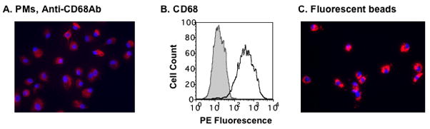
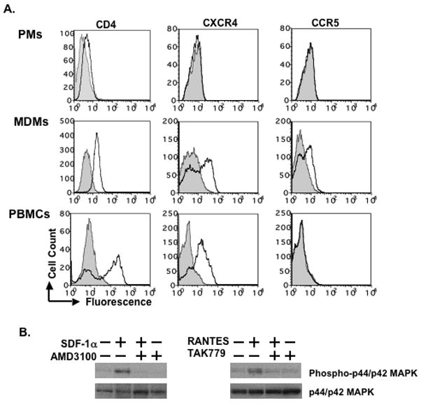
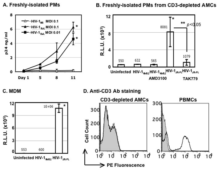
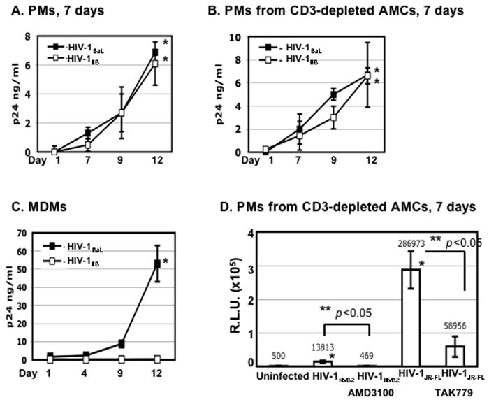
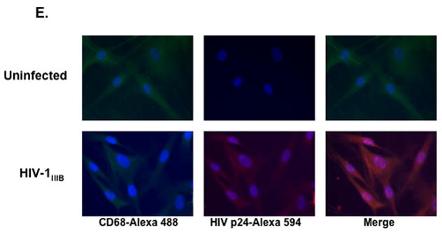
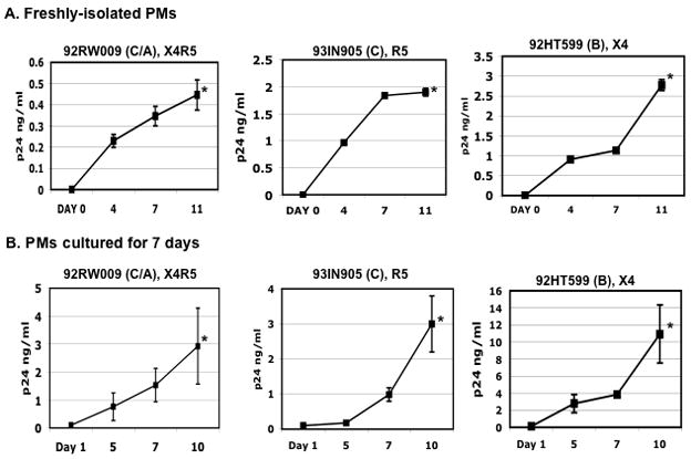
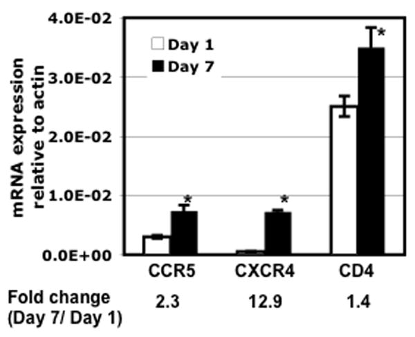
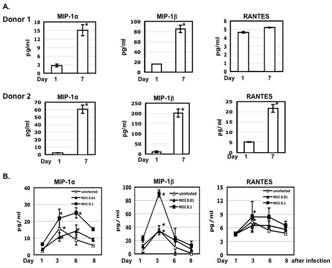
Macrophages are major HIV target cells. They support both productive and latent HIV-1 infection. Susceptibility of primary macrophages to HIV depends on the anatomical location and activation state of the cells. We demonstrate that peritoneal macrophages (PMs) are abundant in ascitic fluid of patients with liver cirrhosis and are susceptible to HIV-1 infection. PMs expressed CD68, a differentiation marker, exhibited phagocytic activity, and survived in culture for 2 months without additional growth factors. Freshly isolated PMs were susceptible to HIV-1 R5 strains but not to X4-T-cell line-adapted strains. Interestingly, after 7 days in culture, PMs acquired susceptibility to X4-T-cell line-adapted strains. HIV entry inhibitors, TAK779 and AMD3100, blocked HIV infection of PMs, indicating that infection by R5 and X4 strains was mediated by CCR5 and CXCR4, respectively. Although PMs did not express detectable cell surface levels of CXCR4 and CCR5, they did express mRNAs of these HIV coreceptors and responded to stimulation by their natural ligands, SDF-1alpha and RANTES. PMs were susceptible to HIV-1 X4, R5, and X4R5 primary isolates. PMs after 7 days in culture produced greater amounts of X4 and X4R5 HIV than freshly isolated PMs. The day-7 PMs were more susceptible to R5 infection in a single-cycle infection assay, but there was no increase in viral production in a multiple-round infection assay. The level of CXCR4 mRNA and production of CC-chemokines (MIP-1alpha, MIP-1beta, and RANTES) increased significantly during 7 days in culture. Our results indicate that PMs are susceptible to receptor-mediated infection by a broad range of HIV strains. These primary macrophages could provide a valuable system for investigating the role of primary macrophages in HIV pathogenesis.


Figures

Figure 1
Figure 1. Peritoneal macrophages (PMs) are highly abundant in ascitic fluid
(A) PMs were prepared by plating AMCs (1–2 × 10⁶ per well) in 6-well plates overnight, followed by washing the cultures four times with PBS. Adherent cells, PMs, were cultured in RPMI with 10% FBS and stained with a mouse monoclonal antibody against CD68, a marker of differentiated macrophages. (B) The expression of CD68 in day-1 PMs was determined by FACS analysis. The gray area represents the signal from cells stained with isotype control antibody, whereas the solid line indicates the signal from cells stained with CD68 antibody. (C) Day-1 PMs phagocytosed Fluoresbrite particles (6.0 mm in diameter). Panels A, and C are 20x magnification. The results represent experiments carried out on cells from three or more patients.
Figure 2
Figure 2. Cell surface expression of CD4, CXCR4 and CCR5 on day-1 PMs
(A) Cell surface expression of CD4, CXCR4 and CCR5 was determined by FACS analysis. MDMs and PBMCs were included as a comparison. The gray area represents the signal from cells stained with isotype control antibody, whereas the solid line indicates the signal from cells stained with CD4, CXCR4 or CCR5 antibody. (B) To examine whether PMs expressed functional CXCR4 or CCR5, cells were exposed to SDF-1α at 300 ng/ml or RANTES at 200 ng/ml for 15 min, respectively. PMs were also pre-treated with or without specific inhibitors of CXCR4 and CCR5 (AMD3100 or TAK779 at 10 μM, respectively) for 1 h followed by stimulation with SDF-1α or RANTES in the presence of inhibitors. Unstimulated PMs were included as controls. Whole cell extracts were prepared and phosphorylation of MAPK (p44/p42) was determined by Western blotting. Blots were then stripped and re-probed with antibodies against p44/p42 MAPK. The results represent three independent experiments using PMs from different patients. Similar data were obtained when day-1 PMs from frozen or freshly-isolated AMCs were used.
Figure 3
Figure 3. Day-1 PMs are susceptible to R5 HIV-1BaL but not to X4 HIV-1IIIB
(A) Freshly-isolated (day-1) PMs were exposed to HIV-1BaL or HIV-1IIIB for 2 h. Unbound virus was removed by washing with PBS, and cells were cultured in RPMI with 10% FBS without additional growth factors. HIV-1 infection was monitored by measuring virus particles in media using an HIV-1 p24 ELISA. The difference in the HIV p24 level from HIV-1BaL-infected cells at day 1 vs day 11 after viral infection was significant (* p< 0.05). (B) Freshly-isolated PMs from CD3-depleted AMCs were exposed to pseudotyped X4 HIV-1HxB2 or R5 HIV-1JR-FL luciferase reporter viruses for 2 h. Cells were washed and cultured for 48 h before measurement of luciferase activity. To determine sensitivity to inhibitors, PMs were treated with entry inhibitors (AMD3100 or TAK-799 at 10μM) for 1 h before viral exposure. Inhibitors were added back to the culture during and after viral exposure. The mean value of luciferase activity is shown. *p< 0.05, uninfected control vs HIV-1JR-FL-exposed cells; **p< 0.05 for HIV-1JR-FL-exposed cells in the presence or absence of TAK-799. (C) MDMs were exposed to pseudotyped X4 HIV-1HxB2 or R5 HIV-1JR-FL luciferase reporter virus and HIV infection was determined at 48h after infection. *p< 0.05, uninfected control vs HIV-1JR-FL-exposed cells. (D) The CD3+ population in AMCs after CD3 depletion was determined by FACS analysis. Freshly-isolated PBMCs were included as a comparison. For experiments in panels A to C, data are mean ± SD of triplicate samples and represent three independent experiments from different donors.
Figure 4
Figure 4. PMs acquire susceptibility to the HIV-1 laboratory X4-TCLA strain
(A and B) PMs from AMCs with or without CD3 depletion were cultured for 7 days before exposure to HIV-1BaL or HIV-1IIIB for 2 h. Unbound virus was removed by washing with PBS, and cells were cultured in RPMI with 10% FBS. HIV-1 infection was determined by HIV-1 p24 ELISA. The difference in the HIV p24 level at day 1 vs day 12 after infection was significant for both strains (*p< 0.05). (C) As a comparison, MDMs were exposed to HIV-1BaL or HIV-1IIIB and HIV production was determined by ELISA. *p<0.05, HIV-1BaL at day 1 vs day 12 after infection. (D) PMs from CD3-depleted AMCs were cultured for 7 days before exposure to pseudotyped X4 HIV-1HxB2 or R5 HIV-1JR-FL luciferase reporter viruses for 2 h. Cells were washed and cultured for 48 h before measurement of luciferase activity. To determine sensitivity to inhibitors, PMs were treated with entry inhibitors (AMD3100 at 10 μM and TAK-799 at 10μM) for 1 h before viral exposure. Inhibitors were added back to the culture during and after viral exposure. The difference in the luciferase activity between uninfected cells and HIV-1HxB2 PMs (*p< 0.05) as well as HIV-infected PMs with or without inhibitors (**p< 0.05) was significant as determined by two-tailed student t test. (E) PMs were cultured for 7 days before exposure to HIV-1IIIB at an MOI 0.1. Unbound virus was removed by washing; cells were cultured for 10 days and stained with antibodies against HIV-1 p24 and CD68 and appropriate secondary antibodies (20x). For experiments in panels A to D, data are the mean±SD of triplicate samples and represent at least three independent experiments using PMs from different patients.
Figure 5
Figure 5. PMs are susceptible to HIV-1 X4, R5, X4R5 primary isolates
Freshly-isolated (day-1 PMs) (Panel A) or day-7 cultures of PMs (Panel B) were exposed to HIV-1 X4, R5 and X4R5 primary isolates for 2 h at 37°C and then unbound virus was removed by washing. The names and tropisms of virus isolates are indicated and viral genotypes are shown in parentheses. HIV-1 production was determined by HIV p24 ELISA. Data are the mean±SD of triplicate samples and represent two independent experiments using peritoneal macrophages from different patients. The difference in the HIV p24 level at day 1 vs day 10 or 11 after infection was significant (*p< 0.05).
Figure 6
Figure 6. CD4, CCR5 and CXCR4 gene expression is up-regulated in PMs after 7 days in culture
Total RNA was prepared from PMs after culturing for 1 or 7 days. The levels of CD4, CCR5, and CXCR4 mRNA were determined by real-time PCR analysis. The results represent three independent experiments using PMs from different patients. The difference in the level of gene expression from cells after culturing for 1 day vs 7 days was significant (*p<0.05).
Figure 7
Figure 7. CC-chemokines are induced in PMs after culture
(A) AMCs (1×10⁶ per well) were plated in a 6-well plate, cultured for 16 h to allow adherence, and washed four times with PBS to remove non-adherent cells. Media from PMs after 1 or 7 days in culture were collected and the level of chemokines were determined by Luminex assays. (B) PMs were prepared by plating AMCs (1×10⁵ cells per well) in a 48-well plate for overnight following by washing. Cells were exposed to R5 HIV-1BaL at an MOI 0.01 or 0.1 for 2 h, washed and cultured in RPMI with 10% FBS. Uninfected cells were included as a comparison. One-half volume of media was collected at the indicated time points to determine the levels of chemokines and HIV p24. Fresh media were added back at each time point. Significant differences between the level of chemokines at day 1 vs day 4 or 7 after culture (panel A) or infection (panel B) are noted (*p< 0.05). The results represented four independent experiments from different patients.

Similar articles

Cited by

References

    1. Cassol E, Alfano M, Biswas P, Poli G. Monocyte-derived macrophages and myeloid cell lines as targets of HIV-1 replication and persistence. J Leukoc Biol Nov. 2006;80(5):1018–1030. - PubMed
    1. Gorry PR, Churchill M, Crowe SM, Cunningham AL, Gabuzda D. Pathogenesis of macrophage tropic HIV-1. Curr HIV Res Jan. 2005;3(1):53–60. - PubMed
    1. Kedzierska K, Crowe SM. The role of monocytes and macrophages in the pathogenesis of HIV-1 infection. Curr Med Chem Nov. 2002;9(21):1893–1903. - PubMed
    1. Clarke JR, White NC, Weber JN. HIV Compartmentalization: Pathogenesis and Clinical Implications. AIDS Rev. 2000;2:15–22.
    1. Igarashi T, Brown CR, Endo Y, et al. Macrophage are the principal reservoir and sustain high virus loads in rhesus macaques after the depletion of CD4+ T cells by a highly pathogenic simian immunodeficiency virus/HIV type 1 chimera (SHIV): Implications for HIV-1 infections of humans. Proc Natl Acad Sci U S A. 2001 Jan 16;98(2):658–663. - PMC - PubMed

Publication types

MeSH terms