

VCAM-1 blockade delays disease onset, reduces disease severity and inflammatory cells in an atopic dermatitis model
- PMID: 20065994
- PMCID: PMC2841723
- DOI: 10.1038/icb.2009.107
VCAM-1 blockade delays disease onset, reduces disease severity and inflammatory cells in an atopic dermatitis model
Abstract
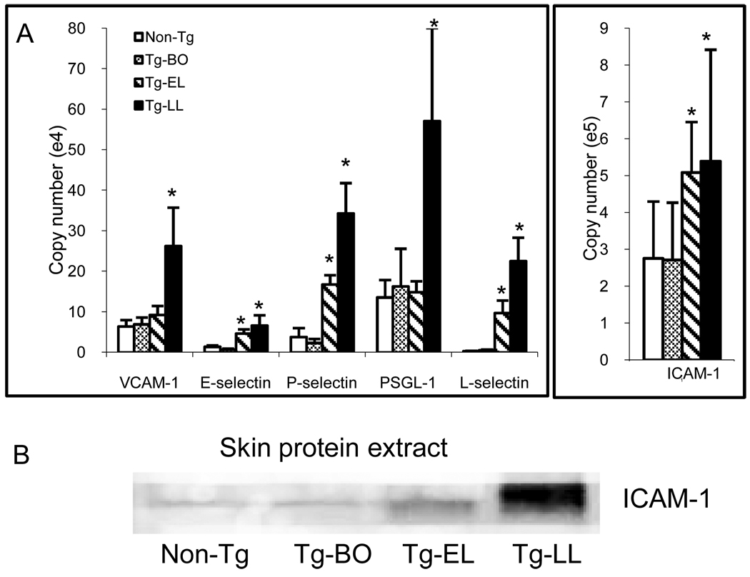
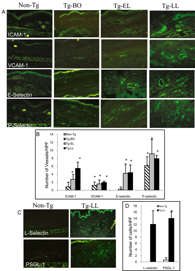
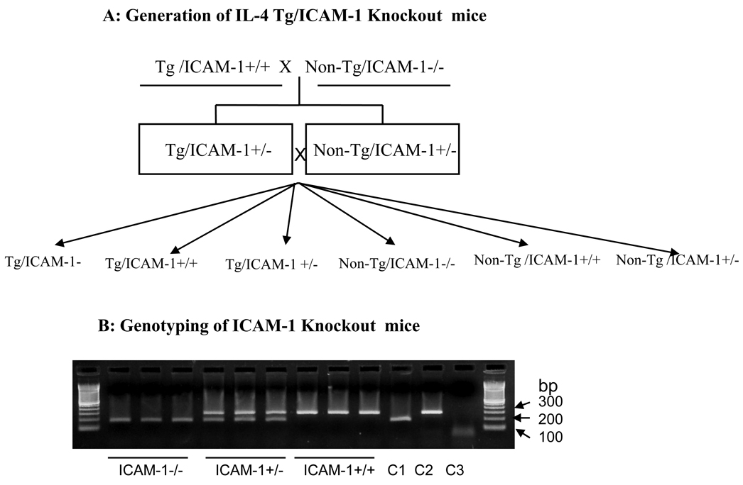
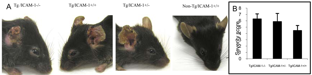
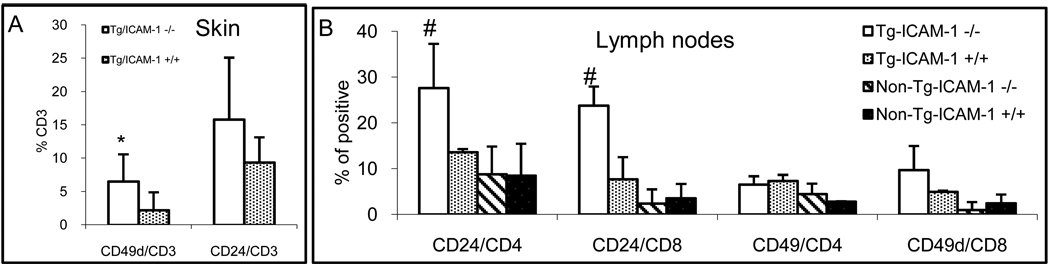
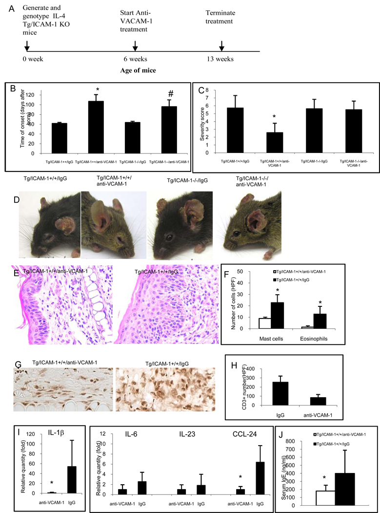
We investigated the functions of critical adhesion molecules ICAM-1 and VCAM-1 in a keratin-14 IL-4-transgenic (Tg) mouse model of atopic dermatitis, the skin lesions of which are characterized by prominent inflammatory cell infiltration, significantly increased mRNAs and proteins of ICAM-1, VCAM-1, E-selectin, P-selectin, L-selectin, and PSGL-1, and significantly increased numbers of dermal vessels expressing these adhesion molecules. We tested the hypotheses that deletion or blockade of these molecules may impede the inflammation by examining the disease progresses in the Tg mice crossed with ICAM-1-knockout mice and Tg mice received anti-VCAM-1-neutralizing antibody. Although the findings of the ICAM-1-knockout Tg mice (Tg/ICAM-1(-/-)) developed skin lesions similar to wide-type ICAM-1 Tg mice (Tg/ICAM-1(+/+)) were surprising, a compensatory mechanism may account for it: the frequency of VCAM-1 ligand, CD49d, on CD3(+) T cells in the lesional skin significantly increased in the Tg/ICAM-1(-/-) mouse, compared with the Tg/ICAM-1(+/+) mice. In contrast, anti-VCAM-1-treated Tg/ICAM-1(-/-) or Tg/ICAM-1(+/+) mice had significantly delayed onset of skin inflammation compared with isotype antibody-treated groups. Moreover, anti-VCAM-1 significantly reduced the skin inflammation severity in Tg/ICAM-1(+/+) mice, accompanied with reduction of mast cell, eosinophil, and CD3(+) T cell infiltration. VCAM-1 is more critical in developing skin inflammation in this model.
Conflict of interest statement
The authors state no conflict of interest.
Figures
Similar articles
-
Expression of VCAM-1, ICAM-1, E-selectin, and P-selectin on endothelium in situ in patients with erythroderma, mycosis fungoides and atopic dermatitis.J Cutan Pathol. 2000 Oct;27(9):436-40. doi: 10.1034/j.1600-0560.2000.027009436.x. J Cutan Pathol. 2000. PMID: 11028813
-
Cell adhesion molecules regulate fibrotic process via Th1/Th2/Th17 cell balance in a bleomycin-induced scleroderma model.J Immunol. 2010 Aug 15;185(4):2502-15. doi: 10.4049/jimmunol.0901778. Epub 2010 Jul 12. J Immunol. 2010. PMID: 20624949 Free PMC article.
-
Adhesion molecules in atopic dermatitis: VCAM-1 and ICAM-1 expression is increased in healthy-appearing skin.Allergy. 1996 Jul;51(7):452-60. Allergy. 1996. PMID: 8863922
-
Endothelial selectins regulate skin wound healing in cooperation with L-selectin and ICAM-1.J Leukoc Biol. 2007 Sep;82(3):519-31. doi: 10.1189/jlb.0307152. Epub 2007 Jun 26. J Leukoc Biol. 2007. PMID: 17595378
-
Skin-homing T cells in human cutaneous allergic inflammation.Immunol Res. 1995;14(4):317-24. doi: 10.1007/BF02935627. Immunol Res. 1995. PMID: 8722046 Review.
Cited by
-
Site-specific targeting of antibody activity in vivo mediated by disease-associated proteases.J Control Release. 2012 Aug 10;161(3):804-12. doi: 10.1016/j.jconrel.2012.05.035. Epub 2012 May 23. J Control Release. 2012. PMID: 22634092 Free PMC article.
-
Forsythia suspensa Suppresses House Dust Mite Extract-Induced Atopic Dermatitis in NC/Nga Mice.PLoS One. 2016 Dec 9;11(12):e0167687. doi: 10.1371/journal.pone.0167687. eCollection 2016. PLoS One. 2016. PMID: 27936051 Free PMC article.
-
Perivascular Adventitial Fibroblast Specialization Accompanies T Cell Retention in the Inflamed Human Dermis.J Immunol. 2019 Jan 1;202(1):56-68. doi: 10.4049/jimmunol.1801209. Epub 2018 Dec 3. J Immunol. 2019. PMID: 30510068 Free PMC article.
-
The involvement of the JAK-STAT signaling pathway in chronic inflammatory skin disease atopic dermatitis.JAKSTAT. 2013 Jul 1;2(3):e24137. doi: 10.4161/jkst.24137. Epub 2013 Aug 15. JAKSTAT. 2013. PMID: 24069552 Free PMC article. Review.
-
Genetic Basis of Irritant Susceptibility in Health Care Workers.J Occup Environ Med. 2016 Aug;58(8):753-9. doi: 10.1097/JOM.0000000000000784. J Occup Environ Med. 2016. PMID: 27206134 Free PMC article.
References
-
- Leung DY, Bieber T. Atopic dermatitis. Lancet. 2003;361(9352):151–160. - PubMed
-
- Cooper KD. Atopic dermatitis: recent trends in pathogenesis and therapy. J Invest Dermatol. 1994;102(1):128–137. - PubMed
-
- Steinhoff M, Steinhoff A, Homey B, Luger TA, Schneider SW. Role of vasculature in atopic dermatitis. J Allergy Clin Immunol. 2006;118(1):190–197. - PubMed
Publication types
MeSH terms
Substances
Grants and funding
LinkOut - more resources
Full Text Sources
Other Literature Sources
Miscellaneous
