

doi: 10.1186/1757-1626-2-9343.
Oral benign fibrous histiocytoma: two case reports
Affiliations
- PMID: 20066060
- PMCID: PMC2804724
- DOI: 10.1186/1757-1626-2-9343
Item in Clipboard
Oral benign fibrous histiocytoma: two case reports
Cases J.
.
Abstract
Fibrous histiocytoma is a benign soft tissue tumour arising as a fibrous mass everywhere in the human body. The involvement of the oral cavity is rare. We report two cases of benign fibrous histiocytoma that localized in the oral cavity. The clinical and histological features of the lesion are reported. Finally, a literature revision of this pathology at the level of the oral cavity is reported.
Figures
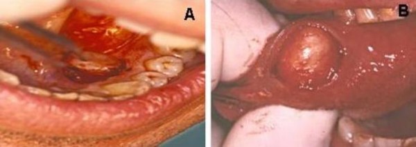
(A) Macroscopical features of Case 1 at surgical excision. (B) Macroscopical features of Case 2 at surgical excision.
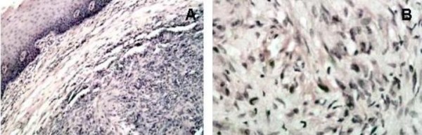
(A) Histological appearance of the lesion in Case 1: the neoplasm is rich in cells of mesenchymal origin; stroma presents myxoid and hyalinization aspects and small foci of necrosis, while fibrous-histiocitic cells displays a storiform or cartwhell pattern (Haemotxylin and Eosin, original magnification ×20). (B) Higher magnification of figure 2A, better showing the cartwhell pattern of the fibrous-histiocitic tumour cells (Haematoxylin and Eosin, original magnification ×40).
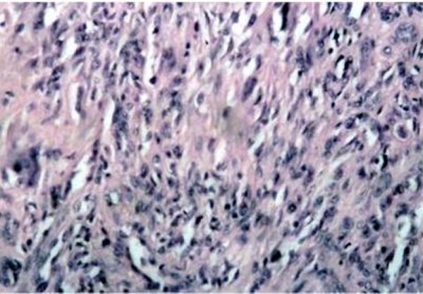
Histological appearance of the lesion in Case 1: the neoplasm consists of a sub-mucosal, cellular aggregation of spindle-shaped, fibroblast-like cells with relatively pale, ovale nuclei; scattered round histiocytic cells are also present (Haemotxylin and Eosin original magnification ×20).
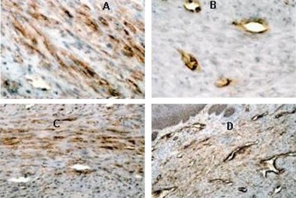
(A) Strong immunohistochemical expression of CD68 in neoplastic cells in Case 1 (ABC, original magnification ×20). (B) Immunohistochemical expression of Factor XIIIa in Case 1: only vessels are positive (ABC, original magnification ×40). (C) Strong immunohistochemical expression of Vimentin in neoplastic cells in Case 2 (ABC, original magnification ×20). (D) Immunohistochemical expression of CD34 in Case 2: only vessels are positive (ABC, original magnification ×20)
References
-
- Fletcher CDM, Unni KK, Mertens F. WHO pathology and genetics of tumours of soft tissue and bone. IARC press Lyon. 2002.
LinkOut - more resources
Full Text Sources
