Review
. 2009 Jul;1(1):a000513.
doi: 10.1101/cshperspect.a000513.

Remodeling epithelial cell organization: transitions between front-rear and apical-basal polarity

Affiliations
Review

Remodeling epithelial cell organization: transitions between front-rear and apical-basal polarity

W James Nelson. Cold Spring Harb Perspect Biol. 2009 Jul.

Abstract

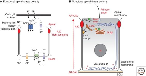
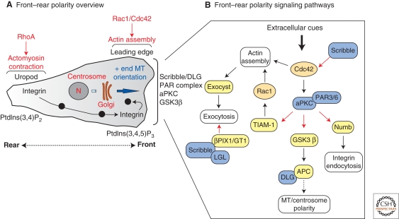
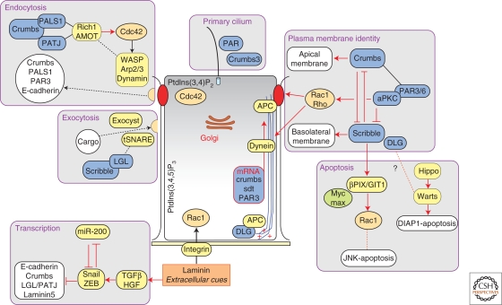
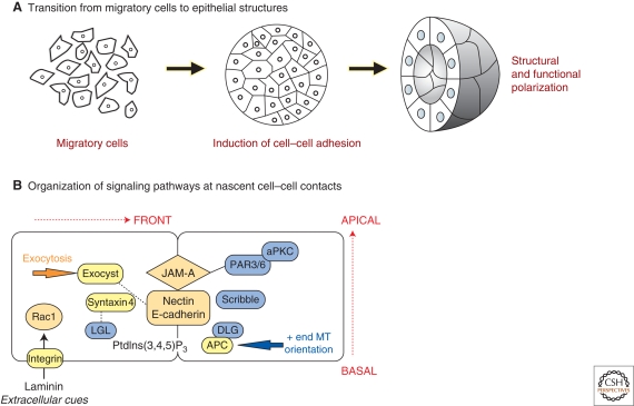
Polarized epithelial cells have a distinctive apical-basal axis of polarity for vectorial transport of ions and solutes across the epithelium. In contrast, migratory mesenchymal cells have a front-rear axis of polarity. During development, mesenchymal cells convert to epithelia by coalescing into aggregates that undergo epithelial differentiation. Signaling networks and protein complexes comprising Rho family GTPases, polarity complexes (Crumbs, PAR, and Scribble), and their downstream effectors, including the cytoskeleton and the endocytic and exocytic vesicle trafficking pathways, together regulate the distributions of plasma membrane and cytoskeletal proteins between front-rear and apical-basal polarity. The challenge is to understand how these regulators and effectors are adapted to regulate symmetry breaking processes that generate cell polarities that are specialized for different cellular activities and functions.


Figures

Figure 1.
Figure 1.
Functional and structural organization of polarized epithelia. (A) Functional apical–basal polarity. Physiological studies of transporting epithelia across the phyla (e.g., crab gill and mammalian kidney nephron) has revealed a remarkable conservation in the distribution of ion channels (Cl channel, K channel) transporters (Na,K,2Cl transporter) and pumps (Na/K-ATPase) between the apical and basolateral plasma membrane domains. The polarized distribution of these proteins generates an apical–basal sodium gradient that is used to move other ions and solutes across the epithelium. (Redrawn and adapted from Cereijido et al. 2004.) (B) Structural apical–basal polarity. Polarized epithelial cells have a distinctive apical–basal polarity in the orientation of cell–cell and cell–extracellular matrix (ECM). Major structures of these cells are also organized in the apical–basal axis: The organization of plasma membrane domains (apical and basolateral), junctional complexes (APC, apical junctional complex), the centrosome (basal body), microtubules and primary cilium, and the secretory pathway (Golgi). For details, see text.
Figure 2.
Figure 2.
Functional and structural organization of migratory mesenchymal cells. (A) Front–rear polarity overview. Migratory cells have a distinctive organization between the front of the cell (leading edge) and rear (uropod). Rho family GTPase activities have different functions in inducing actin polymerization at the leading edge (Cdc42, Rac1), and actomyosin contraction at the rear (RhoA). The centrosome, microtubules, and the secretory pathway (Golgi) are oriented toward the front of the cell. Endocytic and exocytic pathways are also oriented in the rear–front orientation, which allows internalization of integrins from the rear, and exocytosis and endocytosis of integrins in the front, all of which are required for cell migration. Phosphatidylinositides have a polarized distribution in the front–rear polarity. Polarity protein complexes are localized to the front of the cell (Scribble/DLG and PAR/aPKC). (B) Front–rear polarity signaling pathways. Polarity protein complexes (blue) are upstream and downstream of Rho small GTPases (Rac1 and Cdc42) and their downstream effectors (yellow) and end-point effects (white). For details, see text.
Figure 3.
Figure 3.
Functional and structural organization of polarized epithelial cells. Center : Phosphatidylinositides have a distinctive apical–basal polarity; PtdIns(3,4)P2 is localized in the apical membrane, and PtdIns(3,4,5)P3 is localized in the basolateral membrane. Signals from the extracellular matrix (laminin) through integrins and Rac1 activity orient cells, and are required for formation of the apical/luminal domain. Microtubules, which are polarized in the apical–basal axis, are anchored at the basal and lateral membranes (AJC) by APC and other protein complexes; microtubule/dynein-dependent delivery of mRNAs to the apical region is required for proper localization of Crumbs, Sdt (PALS1), and PAR3. Modules: Polarity protein complexes regulate several pathways critical for the establishment and maintenance of apical–basal polarity. Generally, expression of E-cadherin, Polarity protein complexes, and ECM proteins are required to establish and maintain apical–basal polarity (epithelial program). Extracellular cues, including growth factors/cytokines (e.g., TGF-β or HGF) and the transcriptional repressors Snail and ZEB1 down-regulate expression of this epithelial program causing the loss of epithelial differentiation and cell–cell adhesion, and resulting in a front–rear polarization and cell migration (the transcriptional module). Plasma membrane module involves mutually antagonistic regulation of the Crumbs (apical domain) and Scribble (basolateral domain) complexes, and the PAR complex. The Par complex also locally regulates Rac1 and Rho at the AJC (see text for details). Components of the Crumbs (Crumbs3) and PAR complexes are localized to the primary cilium (the primary cilium module) and regulate global cell polarity. The Crumbs complex (the endocytosis module) and Scribble complex (the exocytosis module) control membrane protein organization and stability at the AJC (see text for details). Finally, the Scribble complex appears to play a role in regulating apoptosis (the apoptosis module).
Figure 4.
Figure 4.
Transition between front–rear and apical–basal polarity. (A) Transition from migratory cells to epithelial structures. During development, migratory (mesenchymal) cells coalesce into cell aggregates through cell–cell adhesion, and following induction of the epithelial program, develop into epithelial structures (e.g., a tube). (B) Organization of signaling pathways at nascent cell–cell contacts. Migratory cells interact with each other through their leading edge (front). Cell–cell adhesion is induced by E-cadherin, and additional adhesion proteins (nectin, JAM-A). These adhesion proteins have direct (solid line) and indirect (dotted line) interactions with protein complexes important in apical–basal polarity, including the PAR complex (JAM-A) and the exocyst (directed exocytosis, E-cadherin/nectin); Scribble and LGL are also localized at the leading edge and play critical roles in regulating expocytosis through binding the t-SNARE syntaxin4. Microtubules are oriented toward the front of migrating cells through the localization of the APC (and DLG) complex at the leading edge; APC also plays a role in localizing microtubules to the AJC in the apical–basal axis in polarized epithelial cells.

References

    1. Aigner K, Dampier B, Descovich L, Mikula M, Sultan A, Schreiber M, Mikulits W, Brabletz T, Strand D, Obrist P, et al.2007. The transcription factor ZEB1 (deltaEF1) promotes tumour cell dedifferentiation by repressing master regulators of epithelial polarity. Oncogene 26:6979–6988 - PMC - PubMed
    1. Akhmanova A, Hoogenraad CC, Drabek K, Stepanova T, Dortland B, Verkerk T, Vermeulen W, Burgering BM, De Zeeuw CI, Grosveld F, et al.2001. Clasps are CLIP-115 and -170 associating proteins involved in the regional regulation of microtubule dynamics in motile fibroblasts. Cell 104:923–935 - PubMed
    1. Arimura N, Kaibuchi K 2005. Key regulators in neuronal polarity. Neuron 48:881–884 - PubMed
    1. Audebert S, Navarro C, Nourry C, Chasserot-Golaz S, Lecine P, Bellaiche Y, Dupont JL, Premont RT, Sempere C, Strub JM, et al.2004. Mammalian Scribble forms a tight complex with the betaPIX exchange factor. Curr Biol 14:987–995 - PubMed
    1. Baas AF, Kuipers J, van der Wel NN, Batlle E, Koerten HK, Peters PJ, Clevers HC 2004. Complete polarization of single intestinal epithelial cells upon activation of LKB1 by STRAD. Cell 116:457–466 - PubMed

Publication types

Substances

LinkOut - more resources